Volume 14
-
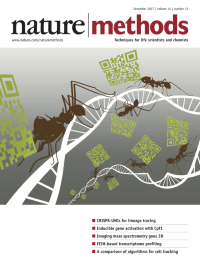
No. 12 December 2017
On the cover: Leaf cutter ants carrying barcodes on DNA symbolize sgRNAs carrying UMIs as part of the DNA-biting CRISPR complex. Image by Izabela Kaminski (http://kaminskigrafik.ch/index.html).
-
No. 11 November 2017
Confocal image of immortalized human skin cells (40× objective) expressing fluorescently tagged keratin. Image acquired by Bram van den Broek in collaboration with Andriy Volkov, Kees Jalink, Reinhard Windoffer and Nicole Schwarz, Netherlands Cancer Institute, Amsterdam. Winner of the 2017 Nikon Small World photomicrography contest (reprinted with permission from B.v.d.B.; image provided by Nikon).
-
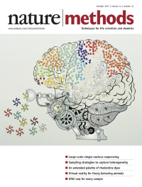
No. 10 October 2017
Droplet microfluidics enable large–scale single–nucleus RNA sequencing of cellular diversity from archived human brain samples. Image by Anna Hupalowska, http://www.annahupalowska.com/.Brief Communication p955.
-
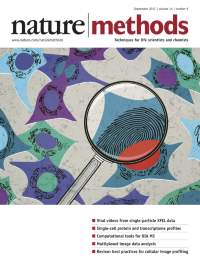
No. 9 September 2017
Computational pipelines have been developed for profiling individual cells from high-throughput image data. Shown is an artistic representation of cell profiling. Cover by Erin Dewalt.Cover image by greyj / iStock / Getty Images Plus. Review p849
-
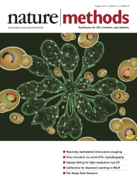
No. 8 August 2017
A massively multiplexed yeast two-hybrid method, CrY2H-seq, enables deep-coverage mapping of the Arabidopsis interactome. Cover prepared by Erin Dewalt, based on design and artwork by Shelly Trigg, Lisa Servilio, and Jamie Simon at The Salk Institute for Biological Studies and by Austin Trigg at Austin Trigg Photography. Article p819
-
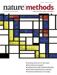
No. 7 July 2017
The image, created by Chiara Nicoletti, was inspired by Hi-C maps and the art of Piet Mondrian. Analysis p679
-
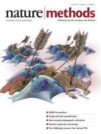
No. 6 June 2017
On the cover: artistic rendering of marker-free coselection in CRISPR editing events. Cover design by Beata Mierzwa, BeataScienceArt.com. Article p615
-
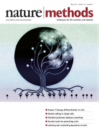
No. 5 May 2017
The cover image represents synergistic signaling in blood progenitors and its effect on cell lineage commitment. Cover prepared by Erin Dewalt, based on an illustration hand drawn by Jennifer Ma, in collaboration with Shreya Shukla and John Edgar, in the laboratory of Peter Zandstra, University of Toronto.
-
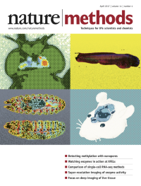
No. 4 April 2017
Pop-art interpretation of specimens imaged with methods highlighted in this issue's Focus on deep imaging of live tissue. Cover by Erin Dewalt based on images provided by T. Katsuki, D. Grover and R. Greenspan (top left); F. Cutrale and L. Trinh (upper right); and R. Chhetri and P. Keller (lower left).
-
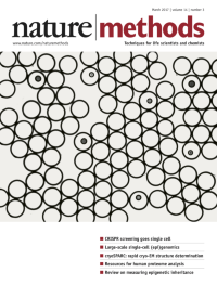
No. 3 March 2017
CROP-seq genetic screening: CRISPR-edited single cells are encapsulated in bead-containing emulsion droplets for single-cell RNA-seq. Cover design by P. Datlinger and N. Winhofer. Article, p297
-
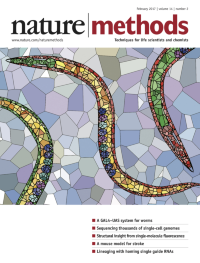
No. 2 February 2017
A modified GAL4–UAS system enables tissue-specific gene expression in the nematode C. elegans, represented artistically by Voronoi tessellation. Artwork by J. Liu, California Institute of Technology. Brief Communication p145.
-
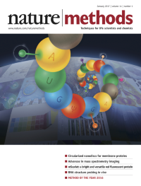
No. 1 January 2017
Epitranscriptome analysis is our Method of the Year for 2016 for its crucial role in understanding RNA modifications on the transcriptome scale. Featured is a stylized image of an RNA with modified bases. Cover design by E. Dewalt.