
Thank you for visiting nature.com. You are using a browser version with limited support for CSS. To obtain the best experience, we recommend you use a more up to date browser (or turn off compatibility mode in Internet Explorer). In the meantime, to ensure continued support, we are displaying the site without styles and JavaScript.

Volume 9 Issue 7, July 2012

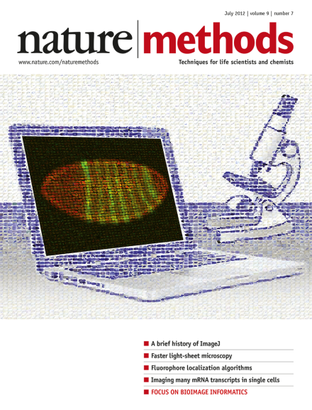
Mosaic image created with the CoverMaker plug-in in Fiji using images of Drosophila gene-expression patterns from histochemistry or fluorescence RNA in situ hybridization experiments. Image courtesy of Pavel Tomancak.

Editorial

  • With the aid of informatics, microscopy is in the midst of a crucial evolution into a more quantitative and powerful technique.

    Editorial

    Advertisement

Top of page ⤴

This Month

Top of page ⤴

Correspondence

Top of page ⤴

Research Highlights

  • A surprising hydrogel formation promises insight on prions and RNA transport.

    • Monya Baker
    Research Highlights
  • Researchers design a simple 'shake & bake' method for the assembly of complex nanostructures from interlocking DNA tiles.

    • Petya V Krasteva
    Research Highlights
  • A nanofluidic device can sort single DNA molecules based on their epigenetic marks.

    • Tal Nawy
    Research Highlights
  • An optical compensation technique enables deeper, crisper two-photon microscopy.

    • Erika Pastrana
    Research Highlights
Top of page ⤴

Methods in Brief

Top of page ⤴

Tools in Brief

Top of page ⤴

Research Highlights

  • Human embryonic stem cells are screened with a panel of antibodies to identify cellular subsets that may correspond to early human progenitor cell populations.

    • Natalie de Souza
    Research Highlights
  • Enzymatic glycosylation and oxidation of methylated DNA together with high-throughput sequencing allows genome-wide base-resolution of 5-hydroxymethyl cytosine distribution.

    • Nicole Rusk
    Research Highlights
Top of page ⤴

Technology Feature

  • To discover new histone marks and interactions, researchers turn to the sophisticated instruments of proteomics.

    • Monya Baker
    Technology Feature
Top of page ⤴

News & Views

  • In toto imaging of living embryos has now become much faster. Light-sheet illumination and fluorescence detection with four objective lenses provide complete coverage of large samples in a snap.

    • Michael Weber
    • Jan Huisken
    News & Views
Top of page ⤴

Commentary

  • Driven by the importance of spatial and physical factors in cellular processes and the size and complexity of modern image data, computational analysis of biological imagery has become a vital emerging sub-discipline of bioinformatics and computer vision.

    • Gene Myers
    Commentary
  • We discuss the advantages and challenges of the open-source strategy in biological image analysis and argue that its full impact will not be realized without better support and recognition of software engineers' contributions to the biological sciences and more support of this development model from funders and institutions.

    • Albert Cardona
    • Pavel Tomancak
    Commentary
  • Bioimaging software developed in a research setting often is not widely used by the scientific community. We suggest that, to maximize both the public's and researchers' investments, usability should be a more highly valued goal. We describe specific characteristics of usability toward which bioimaging software projects should aim.

    • Anne E Carpenter
    • Lee Kamentsky
    • Kevin W Eliceiri
    Commentary
Top of page ⤴

Historical Commentary

  • For the past 25 years NIH Image and ImageJ software have been pioneers as open tools for the analysis of scientific images. We discuss the origins, challenges and solutions of these two programs, and how their history can serve to advise and inform other software projects.

    • Caroline A Schneider
    • Wayne S Rasband
    • Kevin W Eliceiri
    Historical Commentary
Top of page ⤴

Perspective

  • Presented is an overview of the image-analysis software platform Fiji, a distribution of ImageJ that updates the underlying ImageJ architecture and adds modern software design elements to expand the capabilities of the platform and facilitate collaboration between biologists and computer scientists.

    • Johannes Schindelin
    • Ignacio Arganda-Carreras
    • Albert Cardona
    Perspective
  • Icy is a collaborative platform for biological image analysis that extends reproducible research principles by facilitating and stimulating the contribution and sharing of algorithm-based tools and protocols between researchers.

    • Fabrice de Chaumont
    • Stéphane Dallongeville
    • Jean-Christophe Olivo-Marin
    Perspective
Top of page ⤴

Review Article

  • Representative members of the bioimage informatics community review the computational steps and some of the primary software tools available to biologists who are acquiring and analyzing microscopy-based digital image data, with a focus on open-source options.

    • Kevin W Eliceiri
    • Michael R Berthold
    • Anne E Carpenter
    Review Article
Top of page ⤴

Brief Communication

  • The freely available WormToolbox enables high-throughput image analysis of a variety of phenotypes of Caenorhabditis elegans in liquid culture and should prove useful for image-based screens.

    • Carolina Wählby
    • Lee Kamentsky
    • Anne E Carpenter
    Brief Communication
  • The authors describe a method for realigning images from serially sectioned biological specimens that minimizes the effect of artificial deformations in the alignment by applying global elastic constraints. The method is applied to transmission electron microscopy and array tomography image series and is made available through the Fiji platform.

    • Stephan Saalfeld
    • Richard Fetter
    • Pavel Tomancak
    Brief Communication
  • Global optimization of single-molecule localizations using compressed sensing allows stochastic optical reconstruction microscopy (STORM) at high molecular densities and live cell super-resolution imaging with a temporal resolution of 3 seconds.

    • Lei Zhu
    • Wei Zhang
    • Bo Huang
    Brief Communication
  • An analytically exact approach that determines the radial symmetry center of the image of any radially symmetric particle allows faster localization than iterative methods while also giving localization accuracies approaching theoretical limits.

    • Raghuveer Parthasarathy
    Brief Communication
  • Structure determination followed by targeted engineering of the popular photoactivatable fluorescent protein monomeric (m)Eos2 yields mEos3 versions that are more monomeric and less disruptive in protein fusions and also exhibit higher labeling density, brightness and other beneficial properties.

    • Mingshu Zhang
    • Hao Chang
    • Tao Xu
    Brief Communication
  • A selective-plane illumination microscope with two illumination and two detection objectives rapidly records four three-dimensional images of an entire developing fly embryo and processes them into a single high-content image in real time. This allows for cell tracking and quantification of cell shape changes across the embryo. A related paper by Tomer et al. is also in this issue.

    • Uros Krzic
    • Stefan Gunther
    • Lars Hufnagel
    Brief Communication
Top of page ⤴

Article

  • An imaging and registration framework called Virtual Brain Explorer for Zebrafish (ViBE-Z) allows mapping of gene expression patterns and anatomical structures in the zebrafish larval brain. ViBE-Z is provided via a web interface and contains software for image processing, data sets from several developmental stages and a brain atlas.

    • Olaf Ronneberger
    • Kun Liu
    • Wolfgang Driever
    Article
  • Super-resolution microscopy of fluorescently labeled oligonucleotides bound to individual mRNA transcripts is used for highly multiplexed imaging and quantification of transcripts in single cells. The method is used to profile transcripts from 32 stress-response genes in single yeast cells in response to extracellular stress.

    • Eric Lubeck
    • Long Cai
    Article
  • Simultaneous multiview light-sheet microscopy using two illumination and two detection arms with one- or two-photon illumination is coupled to a fast data acquisition framework and analysis pipeline for quantitative imaging and tracking of individual cells and the developing nervous system throughout a living fly embryo. A related paper by Krzic et al. is also in this issue.

    • Raju Tomer
    • Khaled Khairy
    • Philipp J Keller
    Article
Top of page ⤴

Focus

  • Explosive growth in the size and complexity of microscopy-based imaging data and the need to extract more quantitative data from it increasingly requires sophisticated image acquisition and analysis methods and software tools. A collection of articles discusses the role of bioimage informatics in microscopy, specific tools that are available, and the challenges and opportunities in the field.

    Focus
Top of page ⤴

Search

Quick links