Thank you for visiting nature.com. You are using a browser version with limited support for CSS. To obtain
the best experience, we recommend you use a more up to date browser (or turn off compatibility mode in
Internet Explorer). In the meantime, to ensure continued support, we are displaying the site without styles
and JavaScript.
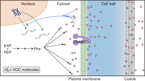
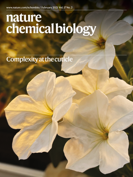
Genetic and chemical perturbations in petunia flowers reveal that the cuticle acts as a sink for volatiles during emission to prevent cellular damage. Alteration of the cuticle affects emission of different volatiles depending on their physiochemical properties.
The plant cuticle has long been considered a barrier to volatile organic compound emission. New research reveals the paradoxical role of the cuticle as a barrier and facilitator of volatile emissions in hybrid petunia.
Yin Yang 1 (YY1) is a transcription factor involved in gene regulation. A recent study has revealed G-quadruplex structures as novel binding targets of YY1, and their interactions regulate DNA looping and gene expression.
Re-channeling metabolic routes in Nicotiana benthamiana allows elucidation and reconstitution of the biosynthesis of the natural products momilactones A and B. Insights into how plants make these and other natural crop protection chemicals promise future biotechnologies for sustainable agriculture.
This Perspective introduces chemical biologists to potentially useful nanotechnologies, aims to inspire nanotechnologists to address questions relevant to chemical biology, and identifies possible opportunities for the two fields to collaborate.
The plant cuticle was initially thought to act as a passive diffusion barrier. Genetic and metabolic analysis reveals that it is also a sink/concentrator for volatiles protecting cells from toxic effects of these hydrophobic compounds.
Qemistree uses fragmentation spectra to predict molecular fingerprints and represent their relationships as a tree, enabling comparison of metabolomics data across different experimental conditions and exploration of chemical diversity in mixtures.
The authors report PROTAC ternary complex structures involving the E3 ligase cIAP1 and target protein BTK, showing that cooperativity is not always correlated with degradation efficiency.
The authors identify the interaction between transcription factor YY1 and DNA G4 structures, which contributes to chromatin looping induced by YY1 dimerization as well as its transcriptional regulation.
Changes in O-GlcNAc levels controlled the actin contraction of fibroblasts in response to sphingosine-1-phosphate (S1P). Specifically, O-GlcNAc modification of the phosphatase MYPT1 maintains its activity to block S1P signaling.
A chemical glycobiology approach reveals that heparan-sulfate glycosaminoglycans regulate vascular development through direct interactions with angiopoietin (Ang) ligands and the Tie1 receptor of the Ang–Tie signaling system.
Native mass spectrometry, HDX-MS and MD simulations define the mechanism for how LPS binding to the Gram-negative outer membrane complex LptDE opens the LptD lateral exit gate and how thanatin impairs transport across the periplasm.
The helical bundle structure of the CC1 domain of STIM1 of the store-activated calcium channel CRAC is crucial to maintaining the channel resting state, and helix–helix interactions can be manipulated to normalize a disease-linked STIM1 mutant.
Redirecting plant diterpene biosynthesis from the chloroplast to the cytosolic, high-flux mevalonate pathway increases intermediate and product titers to support the elucidation and reconstitution of momilactone biosynthesis.
Rather than their expected role in self-immunity, kinases encoded in the biosynthetic gene clusters of nucleoside natural products catalyze the phosphorylation of an early intermediate, a modification that is later removed in a downstream step.
Screening for substrate preference of the SARS-CoV and SARS-CoV-2 main protease Mpro leads to the development of activity-based probes useful for structural analysis and for visualization of active Mpro in infected patient epithelial cells.