
Volume 25 Issue 12, December 2020
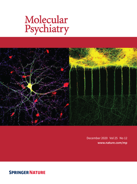
Microfluidic devices elucidate that UPF2-dependent mRNA degradation occurs in dendrites. As exemplified in the left image, neurons (green = GFP, red = GLUR1, blue = SYN1) exhibit complex, intertwined, morphologies, making it difficult to isolate, quantify, and manipulate targets in specific neuronal compartments. In the right image, neurons (green = GFP, red = UPF2) cultured in a custom tripartite microfluidic device allowed Notaras et al. to isolate dendrites from their post-synaptic cell-bodies. In this image, dendrites can be seen penetrating microfluidic device microgrooves to access a middle chamber to synapse with pre-synaptic terminals. This microfluidic device platform allowed Notaras et al. to selectively manipulate the nonsense-mediated mRNA decay pathway in post-synaptic neurons and their dendrites without influencing their reciprocal pre-synaptic neuronal partners. For more information, see the manuscript by Notaras et al. on pages 3360 – 3379.
Image
-
Advertisement
