
Thank you for visiting nature.com. You are using a browser version with limited support for CSS. To obtain the best experience, we recommend you use a more up to date browser (or turn off compatibility mode in Internet Explorer). In the meantime, to ensure continued support, we are displaying the site without styles and JavaScript.

Volume 19 Issue 1, January 2014

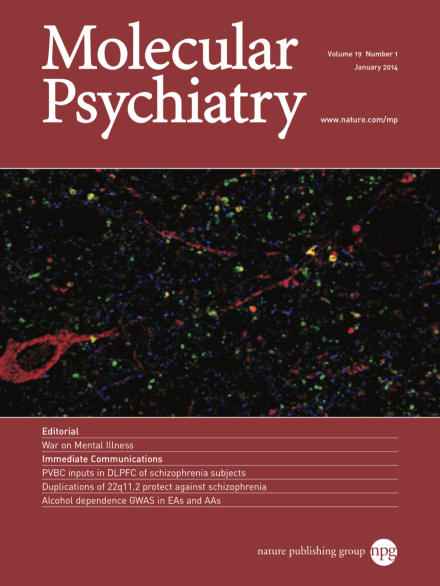
Human dorsolateral prefrontal cortex tissue section triple-labeled for parvalbumin (PV, red), GAD65 (green), and GABAA receptor α1 subunit (blue) illustrating typical neuropil staining. Note the dual-labeled PV/GAD65 axonal boutons (yellow). For more info on this topic, please refer to the article by Glausier et al. on pages 30–36.

Editorial

Top of page ⤴

Image

Top of page ⤴

Letter to the Editor

Top of page ⤴

Review

Top of page ⤴

Immediate Communication

Top of page ⤴

Original Article

Top of page ⤴

Corrigendum

Top of page ⤴

Search

Quick links