Figures, tables and video
From the following article
How to perform esophageal manometry
R. Shaker and C. Hofmann
GI Motility online (2006)
doi:10.1038/gimo90
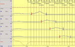
Figure 1
Example of lower esophageal sphincter (LES) rapid pull-through technique (1 cm/s).
Full size figure and legend (76K)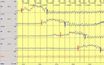
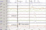
Figure 3
Example of sleeve device properly positioned within the LES.
Full size figure and legend (68K)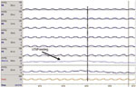
Figure 4
Example of the influence of respiration on esophageal, LES, and gastric pressures.
Full size figure and legend (58K)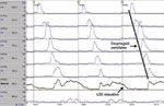
Figure 5
Examples of esophageal peristalsis and LES relaxation during three swallows each of 5 mL of water.
Full size figure and legend (56K)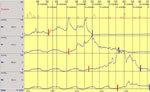
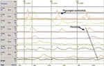
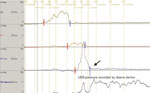
Figure 8
Example of pull-through technique for positioning the sleeve sensor within the UES.
Full size figure and legend (35K)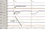
Figure 9
Examples of evaluation of swallow-induced UES relaxation using sleeve device.
Full size figure and legend (55K)