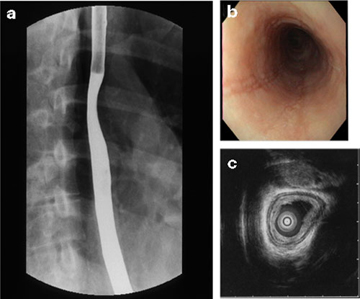
Figure 3 - Radiographic and endoscopic studies from an 18-year-old woman with a longstanding history of dysphagia that began in early childhood.
From the following article
Samuel Nurko and Glenn T. Furuta
GI Motility online (2006)
doi:10.1038/gimo49

a: Barium esophagram showing a small caliber esophagus. b: Endoscopic view of pale mucosa with absent vascular pattern and linear furrows in proximal esophagus. c: Endosonographic image (20 MHz catheter probe) showing thick-walled esophagus with prominent mucosa/submucosa layer and narrow lumen. (Source: Endoscopic ultrasound courtesy of Victor Fox, M.D., Boston. Figure adapted from Fox et al.,3 with permission from the American Society for Gastrointestinal Endoscopy.)
Powerpoint slides for teaching
If the slide opens in your browser, Select "File > Save as" to save it.
Download Power Point slide (977K)
