
Figure 13 - H2O2 in LES epithelial and circular muscle cells.
From the following article
Signal transduction in lower esophageal sphincter circular muscle
Piero Biancani and Karen M. Harnett
GI Motility online (2006)
doi:10.1038/gimo24
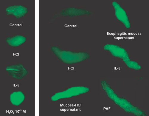
H2O2 levels were examined by confocal microscopy using dihydrorhodamine (DHR 123) as a probe for measurement of intracellular H2O2. Left panel: In untreated epithelial cells (control) H2O2 levels were low and did not change in response to HCl or IL-6. An H2O2-treated epithelial cell is shown as a positive control. H2O2 is membrane permeable, and in the H2O2-treated cell, it penetrates the cytoplasm, demonstrating the adequacy of the technique in detecting cytoplasmic H2O2. Right panel: In normal LES smooth muscle cells (control) H2O2 levels were low and were not affected by HCl. Treatment with supernatant of HCl-filled mucosal sac (mucosa-HCl supernatant), or with supernatant of mucosa from esophagitis animals (esophagitis mucosa supernatant), increased H2O2 in muscle cells. Similarly, treatment with IL-6 or platelet-activating factor (PAF) caused an increase in cytoplasmic H2O2 (Source: Cheng et al.,105 with permission from the American Physiological Society.)
Powerpoint slides for teaching
If the slide opens in your browser, Select "File > Save as" to save it.
Download Power Point slide (490K)