
Figure 6 - Histopathology of achalasia.
From the following article
Pathophysiology of achalasia and diffuse esophageal spasm
Ikuo Hirano
GI Motility online (2006)
doi:10.1038/gimo22
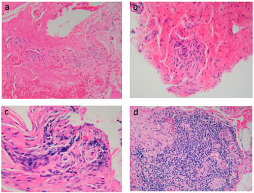
a: Normal myenteric plexus demonstrating multiple ganglion cells and minimal lymphocytic infiltration. b: Mild myenteric inflammation. There is mild lymphocytic inflammation, and ganglion cells can be identified. c: Moderate myenteric inflammation with lymphocytic infiltrate is present. Ganglion cells are absent. d: Severe myenteric inflammation with lymphocytes densely clustered within this myenteric plexus. Ganglion cells are absent. (Source: Hirano and Kahrilas112 with permission from Blackwell Publishing.)
Powerpoint slides for teaching
If the slide opens in your browser, Select "File > Save as" to save it.
Download Power Point slide (1,462K)