Volume 204
-
No. 12 28 June 2008
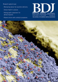
Coloured scanning electron micrograph of the surface of a human tooth (yellow), showing a carpet of spherical bacteria (blue) and red blood cells (red). More than 300 species of bacteria inhabit the human mouth. They feed on the organic film of saliva and food debris that coats the teeth. They produce the enzyme lactate dehydrogenase, which converts sugars into lactic acid. This has a demineralising effect on the tooth enamel, causing tooth caries. The red blood cells may have arisen from bleeding gums, an early sign of gingivitis. COVER IMAGE copyright THIERRY BERROD, MONA LISA PRODUCTION/SCIENCE PHOTO LIBRARY
-
No. 11 14 June 2008
Coloured scanning electron micrograph of a cavity in a tooth caused by caries. The cavity is seen as a depression (at centre) encrusted with brown flaky plaque. The tooth surface is white. Magnifi cation: x150 at 6 x 6 cm size. COVER IMAGE copyright EYE OF SCIENCE/SCIENCE PHOTO LIBRARY
-
No. 10 24 May 2008
Coloured scanning electron micrograph of plaque on the surface of a tooth. Plaque appears as a yellow coating over the grey enamel of the tooth. Magnification: x70 at 6 x 6 cm size. COVER IMAGE copyright DR TONY BRAIN/SCIENCE PHOTO LIBRARY
-
No. 9 10 May 2008
Toothpaste, coloured scanning electron micrograph. COVER IMAGE copyright SUSUMU NISHINAGA/SCIENCE PHOTO LIBRARY
-
No. 8 26 April 2008
Coloured scanning electron micrograph (SEM) of dentine tooth tissue. The holes in the dentine are dental tubules (or canaliculi) formed by the cytoplasmic extensions (green) of odontoblast cells. Magnification: x425 when printed at 10 cm wide. COVER IMAGE copyright STEVE GSCHMEISSNER/SCIENCE PHOTO LIBRARY
-
No. 7 12 April 2008
Coloured scanning electron micrograph (SEM) of a bristle from a used toothbrush, covered in dental plaque. COVER IMAGE copyright STEVE GSCHMEISSNER/SCIENCE PHOTO LIBRARY
-
No. 6 22 March 2008
Light micrograph of a horizontal section through a premolar tooth. The slit-like root canal (centre) is surrounded by a dumb-bell-shaped layer of hard, mineralised dentine (black and white). Covering the dentine is a thin layer of cementum. This is attached to the gums and jaw by the periodontal ligament (light yellow). Magnification: x5 at 6 x 7 cm size. COVER IMAGE copyright INNERSPACE IMAGING/SCIENCE PHOTO LIBRARY
-
No. 5 8 March 2008
"Coloured scanning electron micrograph (SEM) of bacteria and plaque particles found in a decaying tooth. Yellow and green bacteria are seen amongst brown plaque." Magnification: x1,500 at 6 x 6 cm size. COVER IMAGE copyright EYE OF SCIENCE/SCIENCE PHOTO LIBRARY
-
No. 4 23 February 2008
"Coloured scanning electron micrograph (SEM) of a tooth (green) with a dental crown (light blue). The tooth and ceramic crown are coated in debris (orange) which became attached to the tooth when it was extracted for this SEM. Magnification: x10 at 6 x 7 cm size." COVER IMAGE copyright VOLKER STEGER/SCIENCE PHOTO LIBRARY
-
No. 3 9 February 2008
'Coloured scanning electron micrograph showing fl uorapatite crystals at various growth stages.'. COVER IMAGE (C) EYE OF SCIENCE/SCIENCE PHOTO LIBRARY
-
No. 2 26 January 2008
'Coloured scanning electron micrograph (SEM) of a freeze fracture through tooth enamel. The enamel is the outer covering of the crown (visible part) of the tooth. It is the hardest substance in the human body. It is composed of rows of calcium phosphorous salts (light brown) embedded in a protein matrix (darker brown).' COVER IMAGE STEVE GSCHMEISSNER/SCIENCE PHOTO LIBRARY
-
No. 1 12 January 2008
'Light micrograph of a section through a tooth. At the centre of the tooth is the dental pulp (white), which contains the blood vessels and nerves. The majority of the tooth consists of dentine (purple), which is mineralised connective tissue. The tooth is covered by a coat of white enamel (dark purple), the hardest tissue in the human body.'. COVER IMAGE ASTRID & HANNS-FRIEDER MICHLER/SCIENCE PHOTO LIBRARY