Confocal microscopy is a powerful and versatile technique for studying biological phenomena on scales ranging from tissue-level structures to subcellular details. Conducting microscopy experiments with living samples often yields valuable, physiologically relevant data.
However, acquiring clear images can be challenging due to factors such as sample photosensitivity, the relatively fast timescales of biological processes, and the inescapable diffraction limit of light microscopy. Inadequate image quality prevents subsequent analysis and can ultimately hinder research.
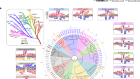
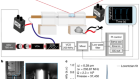
Image scanning microscopy (ISM), an alternative to traditional confocal microscopy, was developed to enhance image resolution and contrast to obtain a clearer picture of any given sample. The technique hinges on the acquisition of additional spatial information about the sample, which enables direct improvements to image quality without detrimental increases in laser illumination power or acquisition time.
This webcast will describe the implementation and advantages of ISM using an array of highly sensitive single photon avalanche photodiodes (SPADs).
Learn:
• How image scanning microscopy (ISM) enhances image quality without compromising other parameters like acquisition time or illumination intensity
• Which applications are particularly well-suited for ISM
• Unique advantages of conducting ISM with a photon-counting detector array
Unable to join the live event? Watch on demand. Register now to ensure that you receive information on how to gain access after the live event.
This webcast has been produced by Nikon Instruments Inc., who retails sole responsibility for content. About this content.
Speakers
Alberto Diaspro, Professor of Applied Physics, Department of Physics, Genoa University

Alberto Diaspro, PhD, is Full Professor of Applied Physics at the Department of Physics of Genoa University (UNIGE), Research Director in Nanoscopy at the Istituto Italiano di Tecnologia (IIT), Affiliate researcher at the Institute of Biophysics (IBF) of the National Research Council (CNR), and Full Academic of the Ligurian Academy of Sciences and Humanities. In 2014 Toshiuki Masai, President Nikon Instruments, Japan, designated Alberto as Director of the Nikon Imaging Centerat IIT. Alberto has provided original and innovative contributions to the development and application of optical microscopy in the linear, non-linear,and super-resolution regimes and has had a critical impact on cellular and molecular biophysics.
Giuseppe Vicidomini, Senior Researcher, Istituto Italiano di Tecnologia (IIT)

Giuseppe Vicidomini studied computer science at the University of Genoa, Italy. He received his PhD in image processing and analysis for fluorescence microscopy from the Laboratory of Advanced Microscopy and Spectroscopy, University of Genoa. As a postdoctoral fellow at the Department of NanoBiophotonics, MaxPlanck Institute for Biophysical Chemistry, Germany, he developed a new approach, based on the temporal analysis of the fluorescence signal, that allows stimulated emission deletion (STED) microscopy to achieve spatial resolution in the tens of nanometers with a substantial reduction of the dose of light required, enabling the effective application of STED microscopy with fluorescent proteins and living cells. He is now principal investigator of the Molecular Microscopy and Spectroscopy research line in the Department of NanoPhysics at the Istituto Italiano di Tecnologia (IIT).
Paolo Bianchini, Scientist, Istituto Italiano di Tecnologia (IIT)

Paolo Bianchini, PhD, studies the biophysical properties of biological molecules and macromolecules both in situ and in vitro utilizing minimally perturbative instrumentation and nanostructured model systems. So far,his research deals with the design, realization, and utilization of biophysical instrumentation such as conventional, confocal, and multi-photon fluorescence microscopies, SHG imaging microscopy, FLIM, single molecule imaging, and STED nanoscopy. He recently applied STED nanoscopy to the quantitative study of single molecule diffusion in living cells. Moreover, he is in charge of the scientific management of the Nikon Imaging Center at the Istituto Italiano di Tecnologia (IIT) and responsible for the partnership with the Nikon Instruments Japan R&D team.
Moderator
Sarah Hiddleston, Nature Research Custom Media

Sarah Hiddleston is a freelance journalist who has worked with Nature Research Custom Media since 2015. Previously, Sarah worked for a decade in Madras (Chennai), India, specialising in health, pharmaceutical and environmental stories. Sarah holds an MA in Investigative Journalism from City University London, an MSc in Political Theory from the London School of Economics, and an undergraduate degree in History from the University of Cambridge, UK.