Volume 13
-
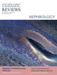
No. 12 December 2017
MUSE (microscopy with UV surface excitation) image of fixed unsectioned kidney, showing a renal artery with elastic lamina surrounded by collagen with renal tubules on either side. Cover image supplied by Richard Levenson, Department of Pathology and Laboratory Medicine, University of California Davis Medical Center at Sacramento, California, USA.
-
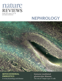
No. 10 October 2017
MUSE (microscopy with UV surface excitation) image of fixed unsectioned kidney, showing a renal artery with elastic lamina surrounded by collagen with renal tubules on either side. Cover image supplied by Richard Levenson, Department of Pathology and Laboratory Medicine, University of California Davis Medical Center at Sacramento, California, USA.
-
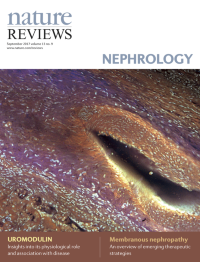
No. 9 September 2017
MUSE (microscopy with UV surface excitation) image of fixed unsectioned kidney, showing a renal artery with elastic lamina surrounded by collagen with renal tubules on either side. Cover image supplied by Richard Levenson, Department of Pathology and Laboratory Medicine, University of California Davis Medical Center at Sacramento, California, USA.
-
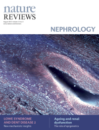
No. 8 August 2017
MUSE (microscopy with UV surface excitation) image of fixed unsectioned kidney, showing a renal artery with elastic lamina surrounded by collagen with renal tubules on either side. Cover image supplied by Richard Levenson, Department of Pathology and Laboratory Medicine, University of California Davis Medical Center at Sacramento, California, USA.
-
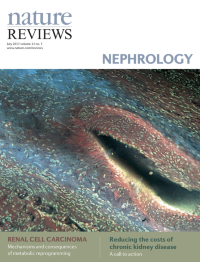
No. 7 July 2017
MUSE (microscopy with UV surface excitation) image of fixed unsectioned kidney, showing a renal artery with elastic lamina surrounded by collagen with renal tubules on either side. Cover image supplied by Richard Levenson, Department of Pathology and Laboratory Medicine, University of California Davis Medical Center at Sacramento, California, USA.
-
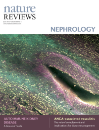
No. 6 June 2017
MUSE (microscopy with UV surface excitation) image of fixed unsectioned kidney, showing a renal artery with elastic lamina surrounded by collagen with renal tubules on either side. Cover image supplied by Richard Levenson, Department of Pathology and Laboratory Medicine, University of California Davis Medical Center at Sacramento, California, USA.
-
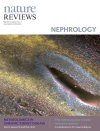
No. 5 May 2017
MUSE (microscopy with UV surface excitation) image of fixed unsectioned kidney, showing a renal artery with elastic lamina surrounded by collagen with renal tubules on either side. Cover image supplied by Richard Levenson, Department of Pathology and Laboratory Medicine, University of California Davis Medical Center at Sacramento, California, USA.
-
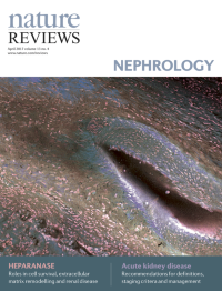
No. 4 April 2017
MUSE (microscopy with UV surface excitation) image of fixed unsectioned kidney, showing a renal artery with elastic lamina surrounded by collagen with renal tubules on either side. Cover image supplied by Richard Levenson, Department of Pathology and Laboratory Medicine, University of California Davis Medical Center at Sacramento, California, USA.
-
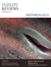
No. 3 March 2017
MUSE (microscopy with UV surface excitation) image of fixed unsectioned kidney, showing a renal artery with elastic lamina surrounded by collagen with renal tubules on either side. Cover image supplied by Richard Levenson, Department of Pathology and Laboratory Medicine, University of California Davis Medical Center at Sacramento, California, USA.
-
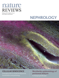
No. 2 February 2017
MUSE (microscopy with UV surface excitation) image of fixed unsectioned kidney, showing a renal artery with elastic lamina surrounded by collagen with renal tubules on either side. Cover image supplied by Richard Levenson, Department of Pathology and Laboratory Medicine, University of California Davis Medical Center at Sacramento, California, USA.
-
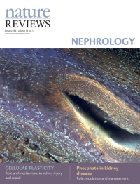
No. 1 January 2017
MUSE (microscopy with UV surface excitation) image of fixed unsectioned kidney, showing a renal artery with elastic lamina surrounded by collagen with renal tubules on either side. Cover image supplied by Richard Levenson, Department of Pathology and Laboratory Medicine, University of California Davis Medical Center at Sacramento, California, USA.