Volume 11
-
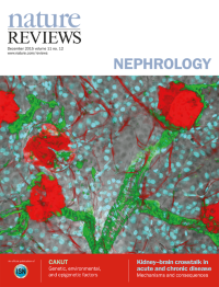
No. 12 December 2015
Cover image supplied by Shih-Jung Peng and Shiue-Cheng Tang, Institute of Biotechnology, Department of Medical Science, National Tsing Hua University, Taiwan. Projection of mouse renal pericytes and their association with glomeruli. The vessel-painted kidney is labelled with the pericyte marker NG2 and imaged by deep-tissue confocal microscopy to illustrate the morphology of renal pericytes. Original lens magnification 25x.
-
No. 11 November 2015
Cover image supplied by Shih-Jung Peng and Shiue-Cheng Tang, Institute of Biotechnology, Department of Medical Science, National Tsing Hua University, Taiwan. Projection of mouse renal pericytes and their association with glomeruli. The vessel-painted kidney is labelled with the pericyte marker NG2 and imaged by deep-tissue confocal microscopy to illustrate the morphology of renal pericytes. Original lens magnification 25x.
Focus
-
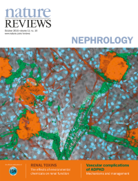
No. 10 October 2015
Cover image supplied by Shih-Jung Peng and Shiue-Cheng Tang, Institute of Biotechnology, Department of Medical Science, National Tsing Hua University, Taiwan. Projection of mouse renal pericytes and their association with glomeruli. The vessel-painted kidney is labelled with the pericyte marker NG2 and imaged by deep-tissue confocal microscopy to illustrate the morphology of renal pericytes. Original lens magnification 25x.
-
No. 9 September 2015
Cover image supplied by Shih-Jung Peng and Shiue-Cheng Tang, Institute of Biotechnology, Department of Medical Science, National Tsing Hua University, Taiwan. Projection of mouse renal pericytes and their association with glomeruli. The vessel-painted kidney is labelled with the pericyte marker NG2 and imaged by deep-tissue confocal microscopy to illustrate the morphology of renal pericytes. Original lens magnification 25x.
-
No. 8 August 2015
Cover image supplied by Shih-Jung Peng and Shiue-Cheng Tang, Institute of Biotechnology, Department of Medical Science, National Tsing Hua University, Taiwan. Projection of mouse renal pericytes and their association with glomeruli. The vessel-painted kidney is labelled with the pericyte marker NG2 and imaged by deep-tissue confocal microscopy to illustrate the morphology of renal pericytes. Original lens magnification 25x.
-
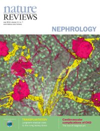
No. 7 July 2015
Cover image supplied by Shih-Jung Peng and Shiue-Cheng Tang, Institute of Biotechnology, Department of Medical Science, National Tsing Hua University, Taiwan. Projection of mouse renal pericytes and their association with glomeruli. The vessel-painted kidney is labelled with the pericyte marker NG2 and imaged by deep-tissue confocal microscopy to illustrate the morphology of renal pericytes. Original lens magnification 25x.
-
No. 6 June 2015
Cover image supplied by Shih-Jung Peng and Shiue-Cheng Tang, Institute of Biotechnology, Department of Medical Science, National Tsing Hua University, Taiwan. Projection of mouse renal pericytes and their association with glomeruli. The vessel-painted kidney is labelled with the pericyte marker NG2 and imaged by deep-tissue confocal microscopy to illustrate the morphology of renal pericytes. Original lens magnification 25x.
-
No. 5 May 2015
Cover image supplied by Shih-Jung Peng and Shiue-Cheng Tang, Institute of Biotechnology, Department of Medical Science, National Tsing Hua University, Taiwan. Projection of mouse renal pericytes and their association with glomeruli. The vessel-painted kidney is labelled with the pericyte marker NG2 and imaged by deep-tissue confocal microscopy to illustrate the morphology of renal pericytes. Original lens magnification 25x.
-
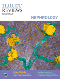
No. 4 April 2015
Cover image supplied by Shih-Jung Peng and Shiue-Cheng Tang, Institute of Biotechnology, Department of Medical Science, National Tsing Hua University, Taiwan. Projection of mouse renal pericytes and their association with glomeruli. The vessel-painted kidney is labelled with the pericyte marker NG2 and imaged by deep-tissue confocal microscopy to illustrate the morphology of renal pericytes. Original lens magnification 25x.
-
No. 3 March 2015
Cover image supplied by Shih-Jung Peng and Shiue-Cheng Tang, Institute of Biotechnology, Department of Medical Science, National Tsing Hua University, Taiwan. Projection of mouse renal pericytes and their association with glomeruli. The vessel-painted kidney is labelled with the pericyte marker NG2 and imaged by deep-tissue confocal microscopy to illustrate the morphology of renal pericytes. Original lens magnification 25x.
-
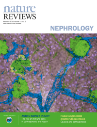
No. 2 February 2015
Cover image supplied by Shih-Jung Peng and Shiue-Cheng Tang, Institute of Biotechnology, Department of Medical Science, National Tsing Hua University, Taiwan. Projection of mouse renal pericytes and their association with glomeruli. The vessel-painted kidney is labelled with the pericyte marker NG2 and imaged by deep-tissue confocal microscopy to illustrate the morphology of renal pericytes. Original lens magnification 25x.
-
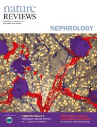
No. 1 January 2015
Cover image supplied by Shih-Jung Peng and Shiue-Cheng Tang, Institute of Biotechnology, Department of Medical Science, National Tsing Hua University, Taiwan. Projection of mouse renal pericytes and their association with glomeruli. The vessel-painted kidney is labelled with the pericyte marker NG2 and imaged by deep-tissue confocal microscopy to illustrate the morphology of renal pericytes. Original lens magnification 25x.