Volume 11
-
No. 12 December 2014
Cover image supplied by Montserrat Reyes, Pathology Laboratory, Faculty of Dentistry, University of Chile, Santiago de Chile, Chile. Optical microscopy image of immunohistochemical staining for β-catenin and p53, as potential contributors to metastatic progression, in a histological section of human oral squamous cell carcinoma.
Focus
-
No. 11 November 2014
Cover image supplied by Montserrat Reyes, Pathology Laboratory, Faculty of Dentistry, University of Chile, Santiago de Chile, Chile. Optical microscopy image of immunohistochemical staining for β-catenin and p53, as potential contributors to metastatic progression, in a histological section of human oral squamous cell carcinoma.
Focus
-
No. 10 October 2014
Cover image supplied by Montserrat Reyes, Pathology Laboratory, Faculty of Dentistry, University of Chile, Santiago de Chile, Chile. Optical microscopy image of immunohistochemical staining for β-catenin and p53, as potential contributors to metastatic progression, in a histological section of human oral squamous cell carcinoma.
-
No. 9 September 2014
Cover image supplied by Montserrat Reyes, Pathology Laboratory, Faculty of Dentistry, University of Chile, Santiago de Chile, Chile. Optical microscopy image of immunohistochemical staining for β-catenin and p53, as potential contributors to metastatic progression, in a histological section of human oral squamous cell carcinoma.
-
No. 8 August 2014
Cover image supplied by Montserrat Reyes, Pathology Laboratory, Faculty of Dentistry, University of Chile, Santiago de Chile, Chile. Optical microscopy image of immunohistochemical staining for β-catenin and p53, as potential contributors to metastatic progression, in a histological section of human oral squamous cell carcinoma.
-
No. 7 July 2014
Cover image supplied by Montserrat Reyes, Pathology Laboratory, Faculty of Dentistry, University of Chile, Santiago de Chile, Chile. Optical microscopy image of immunohistochemical staining for β-catenin and p53, as potential contributors to metastatic progression, in a histological section of human oral squamous cell carcinoma.
-
No. 6 June 2014
Cover image supplied by Montserrat Reyes, Pathology Laboratory, Faculty of Dentistry, University of Chile, Santiago de Chile, Chile. Optical microscopy image of immunohistochemical staining for β-catenin and p53, as potential contributors to metastatic progression, in a histological section of human oral squamous cell carcinoma.
Focus
-
No. 5 May 2014
Cover image supplied by Montserrat Reyes, Pathology Laboratory, Faculty of Dentistry, University of Chile, Santiago de Chile, Chile. Optical microscopy image of immunohistochemical staining for â-catenin and p53, as potential contributors to metastatic progression, in a histological section of human oral squamous cell carcinoma.
-
No. 4 April 2014
Cover image supplied by Montserrat Reyes, Pathology Laboratory, Faculty of Dentistry, University of Chile, Santiago de Chile, Chile. Optical microscopy image of immunohistochemical staining for â-catenin and p53, as potential contributors to metastatic progression, in a histological section of human oral squamous cell carcinoma.
-
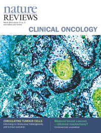
No. 3 March 2014
Cover image supplied by Montserrat Reyes, Pathology Laboratory, Faculty of Dentistry, University of Chile, Santiago de Chile, Chile. Optical microscopy image of immunohistochemical staining for â-catenin and p53, as potential contributors to metastatic progression, in a histological section of human oral squamous cell carcinoma.
-
No. 2 February 2014
Cover image supplied by Montserrat Reyes, Pathology Laboratory, Faculty of Dentistry, University of Chile, Santiago de Chile, Chile. Optical microscopy image of immunohistochemical staining for â-catenin and p53, as potential contributors to metastatic progression, in a histological section of human oral squamous cell carcinoma.
-
No. 1 January 2014
Cover image supplied by Montserrat Reyes, Pathology Laboratory, Faculty of Dentistry, University of Chile, Santiago de Chile, Chile. Optical microscopy image of immunohistochemical staining for â-catenin and p53, as potential contributors to metastatic progression, in a histological section of human oral squamous cell carcinoma.