
Volume 35 Issue 13, December 2010
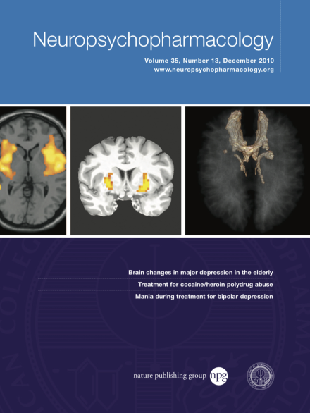
Three dimensional representation of the intersection area of the distal connectivity form historical lesion surgery sites (anterior capsulotomy, anterior cingulotomy, subcaudate tractotomy and limbic leucotomy) as depicted with DTI. The structure in sum shows forceps minor, medial frontal lobe and superolateral medial forebrain bundle. Courtesy of Jan-Christoph Schoene-Bake1, 2, Yaroslav Parpaley1, Bernd Weber1, 2, Jaak Panksepp3, Trevor A Hurwitz4 and Volker A Coenen1 (1University of Bonn Medical Center, Bonn, Germany; 2Life & Brain Center, Bonn, Germany; 3Washington State University, Pullman, WA, USA; 4University of British Columbia, Vancouver, CA, USA).
Commentary
-
Advertisement
