Thank you for visiting nature.com. You are using a browser version with limited support for CSS. To obtain
the best experience, we recommend you use a more up to date browser (or turn off compatibility mode in
Internet Explorer). In the meantime, to ensure continued support, we are displaying the site without styles
and JavaScript.
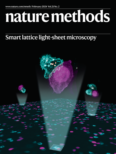
Smart lattice light-sheet microscopy captures rare cellular events. The image shows immune synapses formed between cytotoxic T lymphocytes (cyan) and tumor cells (magenta) within a population of cultured cells. Cytotoxic granules are shown in yellow.
ARTR-seq uses antibody-guided in situ reverse transcription to efficiently and accurately identify RNA-binding protein target sites in as few as 20 cells, or in a formaldehyde-fixed tissue section. The high temporal resolution of ARTR-seq opens opportunities for the investigation of dynamic RNA-binding protein–RNA interactions.
Although single-cell RNA-sequencing has revolutionized biomedical research, exploring cell states from an extracellular vesicle viewpoint has remained elusive. We present an algorithm, SEVtras, that accurately captures signals from small extracellular vesicles and determines source cell-type secretion activity. SEVtras unlocks an extracellular dimension for single-cell analysis with diagnostic potential.
We have developed a framework for the analysis of multi-batch proteome profiling data using isobaric mass tags. Our framework improves quantitative accuracy and increases statistical power by accounting for known sources of variation between batches, thus enabling multiplexed proteome profiling analysis to be performed on large numbers of samples and population cohorts.
This Perspective presents a reliable and comprehensive source of information on pitfalls related to validation metrics in image analysis, with an emphasis on biomedical imaging.
Metrics Reloaded is a comprehensive framework for guiding researchers in the problem-aware selection of metrics for common tasks in biomedical image analysis.
This study shows the importance of proper metrics for comparing algorithms for bioimage segmentation and object detection by exploring the impact of metrics on the relative performance of algorithms in three image analysis competitions.
SnapATAC2 uses a matrix-free spectral embedding algorithm for nonlinear dimension reduction of single-cell omics data, which shows an improved performance in capturing cellular heterogeneity and scalability for large datasets.
DoTA-seq leverages a microfluidic droplet system to isolate and lyse diverse microbes and amplify target genetic loci, enabling high-throughput single-cell sequencing of microbial populations.
This work introduces ARTR-seq for in situ measurement of RNA-binding protein (RBP) binding sites, which has been demonstrated in a small number of cells and for capturing dynamic RBP binding within short timeframes.
CytoCommunity enables both supervised and unsupervised analyses of spatial omics data in order to identify complex tissue cellular neighborhoods based on cell phenotypes and spatial distributions.
The DeepMSA2 pipeline employs iterative alignment search against large genomic and metagenomic sequence databases to construct single- and multichain multiple-sequence alignment (MSA) for proteins. Use of these MSAs shows improvement for deep learning-based protein tertiary and quaternary structure predictions.
smartLLSM uses artificial intelligence-based instrument control to switch between epiflouorescence and lattice light-sheet microscopy to monitor cells at the population level while also capturing multicolor three-dimensional datasets of rare events of interest.
InfraRed-mediated Image Restoration (IR2) uses deep learning to combine the benefits of deep-tissue imaging with NIR probes and the convenience of imaging with GFP for improved time-lapse imaging of embryogenesis.
Content-aware frame interpolation (CAFI) improves the temporal resolution in time-lapse imaging by accurately predicting images in between image pairs. By allowing fewer frames to be imaged, CAFI also enables gentler live-cell imaging.
The thermal-plex method for highly multiplexed imaging uses DNA probes activated when briefly elevated to designated temperatures for rapid, fluidics-free sequential imaging in cells and tissues.
Real-time mid-infrared photothermal imaging of nitrile chameleons enables simultaneous, multiplexed measurement of enzymatic activity in living systems and is poised to reveal the spatiotemporal regulation of enzymes in health and disease.
SynapShot combines ddFPs with engineered synaptic adhesion molecules for real-time observation of the structural plasticity of synapses in cultured cells and animals.