Volume 17
-
No. 12 December 2020
Non-uniform refinement of cryo-EM structuresA reconstruction algorithm called non-uniform refinement accounts for spatial variability in the rigidity and disorder of membrane proteins, yielding improved 3D structures determined from cryo-EM data, as shown here for NaV1.7, a voltage-gated sodium channel.
See Punjani et al.
-
No. 11 November 2020
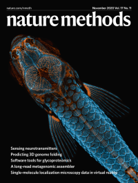
The power of a pictureThe cover photo, the winner of Nikon’s Small World Photomicrography Competition, features a juvenile zebrafish captured using fluorescence labeling, confocal microscopy and image stacking. Bones and scales are labeled in blue, lymphatic vessels in orange.
See Editorial
-
No. 10 October 2020
Behavioral analysis in naturalistic environmentsThe cover photo features a gray mouse lemur (Microcebus murinus), one of the smallest primates and a promising animal model for neuroscience research. The EthoLoop tracking system described in this issue allows analysis of the behavior of these nocturnal, arboreal foragers with unprecedented detail, even in naturalistic environments.
See Nourizonoz et al.
-
No. 9 September 2020
In vivo Brillouin microscopyThe cover depicts a stimulated Brillouin scattering image of the pharynx and the reproductive system of Caenorhabditis elegans, artistically superimposed on a mesh model of the nematode.
See Remer et al.
-
No. 8 August 2020
Red-shifted rhodamines for cellular imagingA photo of quartz cuvettes containing 5 µM solutions of the organic rhodamine dyes Janelia Fluor 479 (JF479), JF559, JF571 and JF593 excited at an angle with a 405-nm laser pointer.
See Grimm et al.
-
No. 7 July 2020
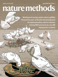
Multilayered scoring system selects sgRNAsThe Vienna Bioactivity CRISPR (VBC) scoring system predicts sgRNAs that efficiently generate loss-of-function alleles. The artwork is inspired by the fairy tale of Cinderella, who receives help from pigeons to sort good and bad peas.
See Michlits et al.
-
No. 6 June 2020
Targeted sequencing perturbationsThe cover illustrates targeted Perturb-seq (TAP-seq), which uses a universal PCR handle and gene-specific primers to amplify genes of interest in single cells. TAP-seq boosts the scale and precision of functional genomics screens and uncovers enhancer–target gene interactions at genome scale.
See Schraivogel et al.
-
No. 5 May 2020
Screening for AAVs with desired tropismsShown is a fluorescence image of mouse brain vasculature. Brain vasculature (cyan) can be genetically targeted with engineered adeno-associated viral capsid AAV-PHP. V1 here carries a fluorescent protein marker (magenta) after systemic injection into the mouse.
See Kumar et al.
-
No. 4 April 2020
Charge detection MS on an OrbitrapThe cover art depicts the process of individual ion mass spectrometry (I2MS), which detects charge of single molecules from electrospray droplets to produce a spectral output directly into the mass domain.
See Kafader et al.
-
No. 3 March 2020
High-speed two-photon microscopyReverberation microscopy image of in vivo mouse brain vasculature. Multiple independent planes at different depths are imaged simultaneously.
See Beaulieu et al.
-
No. 2 February 2020
Protein interaction fingerprinting using deep learningAn artistic representation of molecular surface interaction fingerprinting (MaSIF). Information flows in living systems, such as when proteins interact with each other. The watercolor medium expresses that flow, which is further highlighted by the neural network, identifying their connection.
See Gainza et al.
-
No. 1 January 2020
METHOD OF THE YEAR 2019Our choice for Method of the Year 2019 is single-cell multimodal omics analysis.