Thank you for visiting nature.com. You are using a browser version with limited support for CSS. To obtain
the best experience, we recommend you use a more up to date browser (or turn off compatibility mode in
Internet Explorer). In the meantime, to ensure continued support, we are displaying the site without styles
and JavaScript.
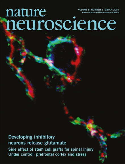
Elimination and strengthening of inhibitory synapses from the medial nucleus of the trapezoid body (MNTB) are essential for the formation of a precise tonotopic map in the lateral superior olive, but the mechanisms behind this plasticity are unclear. Kandler and colleagues now find that these inhibitory MNTB terminals co-release the excitatory transmitter glutamate during the period of synapse elimination, which activates postsynaptic NMDA receptors. Here, an axon terminal from a dye-filled GABA/glycinergic MNTB neuron (red) is immunolabeled against the vesicular glutamate transporter VGLUT3 (blue) and the synaptic vesicle protein SV2 (green). (pp 257 and 332)
How activity-dependent synaptic plasticity shapes the development of inhibitory synapses has remained unclear. In this issue, Gillespie et al. show that in the developing rat auditory system, inhibitory synapses transiently co-release glutamate. The consequent activation of postsynaptic NMDA receptors may be critical for the plasticity mechanisms that determine tonotopic sharpening.
Stem cells transplanted after spinal cord injury mostly generate astrocytes, which can promote aberrant sprouting of sensory neurons, leading to allodynia. Making cells produce more oligodendrocytes reduces allodynia and improves functional recovery.
Having control over a stressful situation can reduce its negative physiological and cognitive consequences. In this issue, a new study in rats suggests that descending inputs from the prefrontal cortex to the serotonergic midbrain signal the controllability of stress.
A recent study of an Olig1 knockout mouse concludes that remyelination after injury may occur by a different mechanism from myelination during normal development, but another report suggests that this mouse model should be interpreted cautiously.
In a technical tour de force, Okhi et al. image the activity of thousands of visual cortical neurons in vivo at a single-cell resolution, and examine their orientation and direction selectivity. Their results show that cortical maps can be built with single-cell precision.