Volume 8
-
No. 12 December 2006
The armadillo protein p0071 regulates Rho signalling during cytokinesis and p0071 overexpression (red) leads to multinucleation.p1432
-
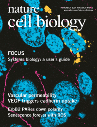
No. 11 November 2006
VE-cadherin endocytosis is promoted by VEGF.
-
No. 10 October 2006
In C. elegans, the yolk (green) provides the oocyte with polyunsaturated fatty acids, which control sperm (red) recruitment to the site of fertilization.p1143
-
No. 9 September 2006
CFTR is important for lysosomal acidification in aveolar macrophages.p933
-
No. 8 August 2006
During ageing of primary mouse embryonic fibroblasts, cyclin D1 (green) relocalizes from the nucleus (blue) to the cytosol(the actin cytoskeleton is shown in red).p877
-
No. 7 July 2006
InsP6 directly modulates Dbp5 in mRNA export. In situ hybridization of cells lacking Nup42 and InsP6 (bottom) or of the same cells expressing a dominant allele of Dbp5 (top). mRNA localization is pseudocolored green, DNA is red.p668
-
No. 6 June 2006
The NuMA-related Mud protein regulates neuroblast spindle orientation. Larval brain showing neuroblast asymmetric division visualized with a GFPmicrotubule marker; single neuroblasts and their progeny are pseudocoloured. Optic lobe, uncoloured.
-
No. 5 May 2006
Defining a hierarchy of novel complexes that associate with centromeric CENP-A nucleosomes. Centromeres stained with anti-centromere antibodies are pseudocoloured and tubulin is shown in white.p458
-
No. 4 April 2006
Rho1 regulates crosslinking of F-actin and microtubules by Capu and Spire. Stabilized microtubules (red) and F-actin (green) were incubated with Capu, Spire and Rho1 proteins.
-
No. 3 March 2006
Myosin ll organizes growth cone cytoplasmic domains. Montage of a neuronal growth cone from Aplysia alifornica permeabilized in cytoskeleton stabilizing buffer before fixation and subsequent F-actin, myosin ll, and microtubule labelling.
-
No. 2 February 2006
T-lymphoblasts follow a transcellular route of diapedesis through human dermal microvascular endothelial cells stimulated with TNF-α. The Tlymphoblast is shown by phalloidin staining (red), and endothelial caveolin-1 and ICAM-1 are shown in green and blue, respectively.p113
-
No. 1 January 2006
Fly pupal retina with all cells overexpressing Expanded, depicting the overproliferation phenotype of warts mutant cells (lacking green staining; cell outlines in blue, photoreceptor nuclei in red/orange/green). The 'Plastic Wrap' filter from Adobe Photoshop was applied to this image.