
Thank you for visiting nature.com. You are using a browser version with limited support for CSS. To obtain the best experience, we recommend you use a more up to date browser (or turn off compatibility mode in Internet Explorer). In the meantime, to ensure continued support, we are displaying the site without styles and JavaScript.

Volume 34 Issue 12, December 2016

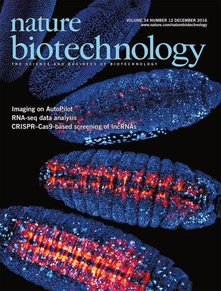
High-resolution, live imaging of an entire developing Drosophila embryo at different stages, with adaptive light-sheet microscopy, highlighting the nuclei of all of the embryo's cells in blue and its developing nervous system in red and yellow. Royer et al. present an approach to continuously evaluate changes in the embryo's optical properties and to adapt the microscope's configuration to these dynamic conditions to recover and maintain high spatial resolution (p 1267). Image credit: Julia Kuhl, Loïc A. Royer & Philipp J. Keller

Editorial

  • Despite an increasingly strident outcry against drug prices in the United States, manufacturers likely face an evolution in the reimbursement landscape, rather than a revolution.

    Editorial

    Advertisement

Top of page ⤴

News

Top of page ⤴

Bioentrepreneur

Top of page ⤴

Correspondence

Top of page ⤴

Feature

  • Chasing treatments and gouging patients in a bloated, capitalist healthcare system.

    • Brady Huggett
    Feature
Top of page ⤴

Patents

Top of page ⤴

News & Views

Top of page ⤴

Perspective

  • Modern biological research increasingly relies on image data as a primary source of information in unraveling the cellular and molecular mechanisms of life. The quantity and complexity of the data generated by state-of-the-art microscopes preclude visual or manual analysis and require advanced computational methods to fully explore the wealth of information. In addition to making bioimage analysis more efficient, objective, and reproducible, the use of computers improves the accuracy and sensitivity of the analyses and helps to reveal subtleties that may be unnoticeable to the human eye. Many methods and software tools have already been developed to this end, but there is still a long way to go before biologists can blindly trust automated measurements. Here, we summarize the current state of the art in bioimage analysis and provide a perspective on likely future developments.

    • Erik Meijering
    • Anne E Carpenter
    • Jean-Christophe Olivo-Marin
    Perspective
Top of page ⤴

Analysis

Top of page ⤴

Brief Communication

Top of page ⤴

Article

Top of page ⤴

Letter

Top of page ⤴

Erratum

Top of page ⤴

Careers and Recruitment

Top of page ⤴

Search

Quick links