
Volume 26 Issue 7, July 2021
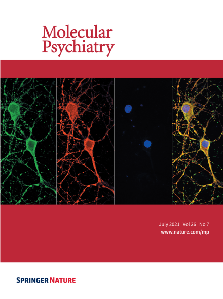
Immunofl uorescent staining of rat primary neurons (DIV. 14). Images show neuronal TrkB and APP co-localized on the cell surface. Neurons are labeled green with APP, red with TrkB, and blue with DAPI. For more information see the article by Yiyuan Xia et al. on page 2943–2963.
Image
-
Advertisement
