Thank you for visiting nature.com. You are using a browser version with limited support for CSS. To obtain
the best experience, we recommend you use a more up to date browser (or turn off compatibility mode in
Internet Explorer). In the meantime, to ensure continued support, we are displaying the site without styles
and JavaScript.
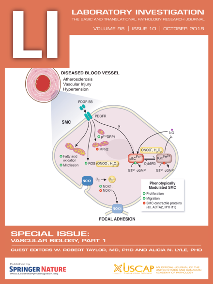
The cover shows a schematic illustration of the mechanisms of smooth muscle cell phenotypic switching. For more information, see the paper by Durgin and Straub, p 1254, this issue
Oxidative stress and smooth muscle cells (SMCs) are major contributors to pulmonary arterial hypertension, atherosclerosis, and systemic hypertension. In this review, the authors discuss how redox balance in SMC by vascular NADPH oxidases, mitochondria bioenergetics, as well as the soluble guanylate cyclase–cyclic guanosine monophosphate–protein kinase G pathway can functionally change SMC from contractile to phenotypically diverse cells and vice versa.
In this study, systemic hemodynamics and both aortic and intrahepatic vascular reactivity in a rat model of severe steatosis were investigated. Portal hypertension and signs of a hyperdynamic circulation were demonstrated. NO-independent, COX-2 mediated extrahepatic arterial hyporeactivity, as well as NO-independent alpha-1-adrenergic and endotheline-1 mediated intrahepatic vascular hyperreactivity are demonstrated, likely contributing to the observed portal hypertension in steatosis.
The authors investigated the effect of therapies based on the JAK/STAT regulator SOCS1 to combat diabetes-induced oxidative stress. The SOCS1 gene and peptidomimetic administration prevents renal damage and atherosclerosis in streptozotocin-diabetic mice by reducing oxidative stress and enhancing antioxidant genes. Mechanistically, SOCS1 prevents activation/expression of NADPH oxidase via PI3K and STAT1 inhibition.
Extracellular vesicles (EVs) comprise a wide range of bilayer membrane-enclosed nanovesicles secreted by nearly all cell types and present in all human body fluids. In dilated cardiomyopathy, which is a frequent cause of heart transplantation, unraveling the specific combinations of cargo proteins within the EVs that peripherally circulate in patients hold promising potential for biomarker discovery.
A single high fat meal can induce pathological red blood cell (RBC) remodeling and oxidative stress, in conjunction with elevations in plasma, RBC-bound myeloperoxidase (MPO) and MPO-mediated high-density lipoprotein oxidation. These findings demonstrate that consumption of heavy meals enriched in fat may promote destabilization of vulnerable plaques leading to acute myocardial infarction.
This paper examines whether inhibitor kappa B kinase 2 (IKK2) acts as a myosin light chain kinase (MLCK) or regulates the activity of myosin light chain phosphatase (MLCP), and therefore affects vasoconstriction. The authors found that IKK2 regulates MLCP activity by phosphorylating the MYPT1 subunit. Furthermore, SC-514, an IKK2 inhibitor, induces relaxation of mesenteric arteries. Therefore, IKK2 may be a new pharmacological target for treatment of various vascular diseases.
Hyaluronan (HA) is known to inhibit osteoblast differentiation, but its importance in vascular calcification needed clarification. In this study, the authors identify HA as a novel negative regulator of osteogenic differentiation of vascular smooth muscle cells via bone morphogenetic protein 2 signaling. These results indicate that HA may be a novel potential therapeutic agent for the treatment of vascular calcification.
Currently, therapeutic choices for patients with pulmonary hypertension (PH) are limited. This paper reveals that dipeptidyl peptidase IV inhibition alleviates pulmonary arterial remodeling in PH by decreasing the proliferation and migration of pulmonary arterial smooth muscle cells by regulating PTEN/AKT/MAPK signaling. Therefore, soluble DPP-4 might be a diagnostic or prognostic marker for PH; and DPP-4 inhibitors, potential treatments.
Blood-brain barrier dysfunction and disruption are significant events in central nervous system inflammatory processes. This study shows that TNF-α and VEGF decrease cell-surface cellular prion protein, an adhesion protein, on human brain microvascular endothelial cells, which are a major component of the blood brain barrier. They also show that loss of cellular prion protein results in decreases of the tight junction proteins claudin-5 and occludin and increases permeability of the endothelial layer.