
Volume 95 Issue 2, February 2015
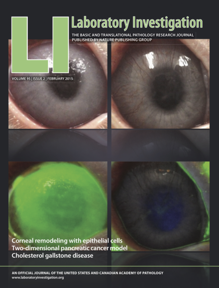
The upper and lower rows show the ocular surfaces of a rabbit corneal deficiency model before and after fluorescein staining, respectively. The left panels are at week 1 and the right panels, week 48. For more information see the paper by Kameishi et al, this issue.
Inside the USCAP Journals
-
Advertisement
