Volume 100
-
No. 12 December 2020
Immunofluorescent images of β-catenin-positive osteoblasts in bone samples from mice 2 and 12 months of age. For more information, see the paper by Yang et al, p 1499, this issue.
-
No. 11 November 2020
The cover shows immunofluorescence of cystic fibrosis pig gallbladder organoids. Blue, green, and gray represent DAPI, Na+/K+ ATPase, and actin, respectively. For more information, see the paper by Zarei et al, p 1388, this issue.
-
No. 10 October 2020
The paper by Roy et al. ( p 1374) shows the use of deep learning for histological assessment of liver biopsies, resulting in accurate quantification of hepatic steatosis. The cover shows visualization of segmented steatosis droplets in masks of distinct colors.
-
No. 9 September 2020
The cover shows medulloblastoma tumoroids. For more information, see the article by Cheng et al, this issue (p 1208).
-
No. 8 August 2020
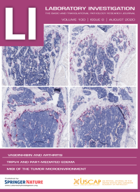
The cover shows the effects of vasoinhibin in murine antigen-induced arthritis. For more information, see the paper by Ortiz et al, p 1068, this issue.
-
No. 7 July 2020
The cover shows monocyte adhesion to an endothelial cell. For more information, see the paper by Posta et al, this issue, p 986.
-
No. 6 June 2020
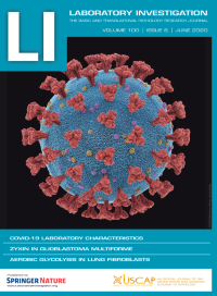
The cover shows a depiction of severe acute respiratory syndrome coronavirus 2 (SARS-CoV-2). Credit: KTSDESIGN/Science Photo Library/Getty. Used with permission.
-
No. 5 May 2020
The paper by Tsuru et al., this issue (p 738) describes how RAMP1 signaling in immune cells regulates inflammation-associated lymphangiogenesis. The cover shows lymphatic vessels in whole-mount diaphragm tissue stained with antibodies specific for Lyve-1 and mediastinal lymph nodes in the pleural cavity after injection of FITC-dextran.
-
No. 4 April 2020
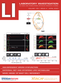
The cover shows localization of extra-mitochondrial citrate synthase in the sperm and its involvement in egg activation. For more information see the paper by Kang et al, this issue.
-
No. 3 March 2020
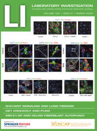
Cover: The cover images are from the paper by Cao et al., which links the Shh-Wnt signaling cascade with the fibrogenic activity of lung resident mesenchymal stem cells and the development of pulmonary fibrogenesis.
-
No. 2 February 2020
The cover depicts the roles of TRPV4 channel in key aspects of cancer progression. For more information, see the paper by So et al, p 199, this issue.
-
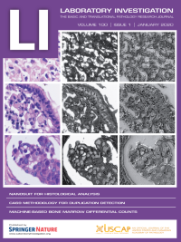
No. 1 January 2020
Cover: The cover shows identification of nuclei in paraffin sections using the NanoSuit method. Upper row, malignant mesothelioma; middle row, gastric carcinoma (signet ring cell carcinoma); and lower row, breast cancer sample. Left column, hematoxylin and eosin (H&E) staining; middle column, low-vacuum scanning electron microscopy images of samples stained with gold (III) chloride, taken in BSE mode; and right column, mixed low-vacuum scanning electron microscopy images of samples stained with gold (III) chloride. For more information, see the paper by Kawasaki et al, this issue (p 161).