Volume 29 Issue 10-11, November 2022
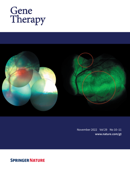
Cover Credit: Sheep fundus following subretinal injection of a modified AAV9 vector. Left: A composite image showing two subretinal blebs immediately after injection. Right: A composite image showing GFP expression 12 weeks after injection. The 7m8 modification of the GFP-carrying vector allowed radial spread of the vector, leading to coalescence of the two original blebs. Such spread has implications on the safety of the subretinal injection, as it potentially enables treating the central retina (or fovea) using a safer, more peripheral subretinal deposition of the vector.