Figures, tables and video
From the following article
Anatomy, development, and physiology of the lungs
Richard M. Effros
GI Motility online (2006)
doi:10.1038/gimo73
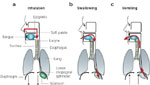
Figure 1
Intersection of respiratory and gastrointestinal (GI) tracts.
Full size figure and legend (26K)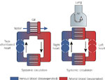
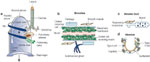
Figure 3
Schematic diagram of lung anatomy with cross-sections of bronchi, bronchioles alveolar ducts, and alveoli.
Full size figure and legend (55K)
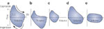
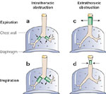
Figure 6
Effects of intrathoracic and extrathoracic obstruction on the caliber of airways.
Full size figure and legend (25K)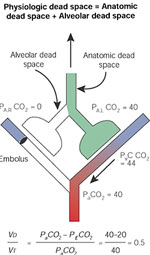
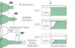
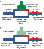
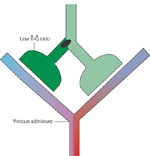
Figure 11
The effect of mismatched ventilation  and perfusion
  on arterial oxygenation.