
Figures, tables and video
From the following article
Endoscopic evaluation of esophageal motility disorders
Susan E. McCormick and Richard A. Kozarek
GI Motility online (2006)
doi:10.1038/gimo29
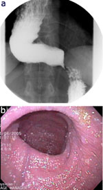
Figure 1
a: Barium esophagram showing a dilated, tortuous esophagus and a "bird's beak" appearance of the lower esophageal sphincter (LES).
Full size figure and legend (18K)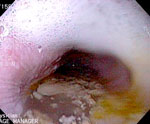
Figure 2
Dilated, fluid-filled esophagus in a patient with achalasia.
Full size figure and legend (65K)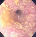
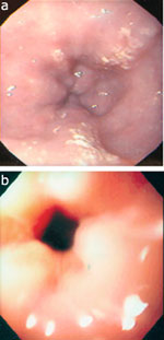
Figure 4
Two examples of a puckered lower esophageal sphincter in patients with achalasia.
Full size figure and legend (16K)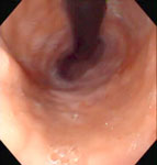
Figure 5
Retroflexed view showing a patulous gastroesophageal junction in a patient with scleroderma.
Full size figure and legend (48K)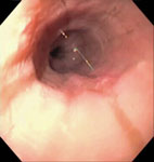
Figure 6
A fairly normal-appearing esophagus in a patient with symptomatic nutcracker esophagus.
Full size figure and legend (52K)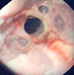
Figure 7
Several epiphrenic diverticula in a patient with reflux esophagitis and a peptic stricture.
Full size figure and legend (64K)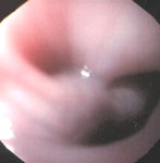
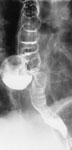
Figure 9
Barium x-ray swallowing study showing a large mid-esophageal pulsion diverticulum.
Full size figure and legend (42K)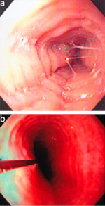
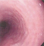
Figure 11
Fine circumferential folds, which disappear with continued air insufflation.
Full size figure and legend (66K)