Figures, tables and video
From the following article
William G. Paterson
GI Motility online (2006)
doi:10.1038/gimo13
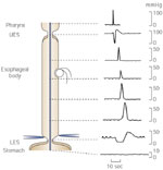
Figure 1
Primary peristalsis as recorded by an intraluminal manometry catheter.
Full size figure and legend (38K)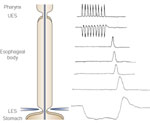
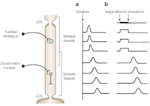
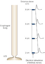
Figure 4
Electrical stimulation of intrinsic nerves in circular smooth muscle strips.
Full size figure and legend (27K)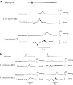
Figure 5
Simultaneous recording of electrical and mechanical activity in opossum smooth muscle esophagus.
Full size figure and legend (33K)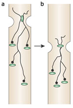
Figure 6
Model showing the marked delay in onset of distal esophageal contractions during peristalsis.
Full size figure and legend (25K)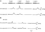
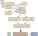
Figure 8
Pathways involved in acetylcholine-induced contraction of esophageal circular smooth muscle.
Full size figure and legend (21K)