
Thank you for visiting nature.com. You are using a browser version with limited support for CSS. To obtain the best experience, we recommend you use a more up to date browser (or turn off compatibility mode in Internet Explorer). In the meantime, to ensure continued support, we are displaying the site without styles and JavaScript.

Volume 204 Issue 6, 22 March 2008

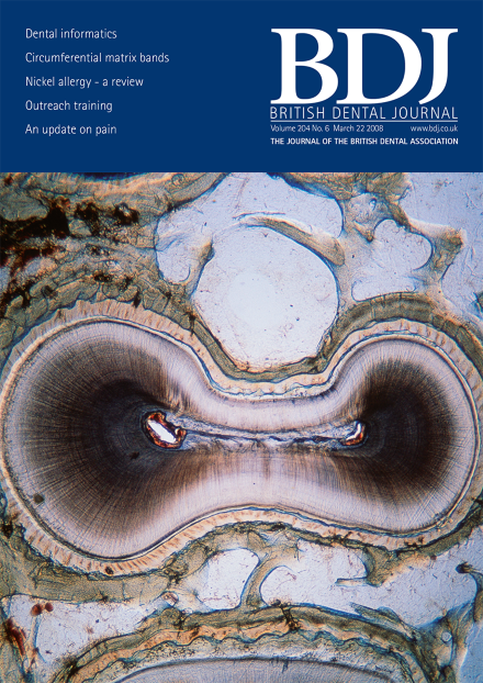
Light micrograph of a horizontal section through a premolar tooth. The slit-like root canal (centre) is surrounded by a dumb-bell-shaped layer of hard, mineralised dentine (black and white). Covering the dentine is a thin layer of cementum. This is attached to the gums and jaw by the periodontal ligament (light yellow). Magnification: x5 at 6 x 7 cm size. COVER IMAGE copyright INNERSPACE IMAGING/SCIENCE PHOTO LIBRARY

Editorial

Top of page ⤴

Letter

Top of page ⤴

News

Top of page ⤴

Practice

Top of page ⤴

Erratum

Top of page ⤴

Practice

Top of page ⤴

Other Journals in Brief

Top of page ⤴

Research Summary

Top of page ⤴

Education

Top of page ⤴

Product News

Top of page ⤴

Conference

  • Professor Renton will be giving 'An update on pain' on Saturday 3 May at the 2008 British Dental Conference and Exhibition, held at the Manchester Central Convention Complex.

    • T. Renton
    Conference
Top of page ⤴

Erratum

Top of page ⤴

Obituary

Top of page ⤴

Special Feature

Top of page ⤴

Research

Top of page ⤴

Search

Quick links