PROVIDED BY

AND

This page has been archived and is no longer updated
In the early 1950s,
experiments by two teams of researchers, Salvador Luria working with Mary Human
and Joe Bertani working with Jean Weigle, showed that some strains of bacteria
were more resistant to viral infections than others. Viruses that infect
bacterial cells are called bacteriophages. Their main goal is to produce more
bacteriophages by injecting their genome into a bacterial host cell, using the
host cell machinery to copy their genome, and expressing bacteriophage genes.
The researchers found, however, that some strains of bacteria appeared to be
less vulnerable to bacteriophage infections than others and resisted the
hijacking of their cell machinery by bacteriophages. A deeper look into the apparent
self-defense mechanisms of these bacteriophage-resistant bacteria revealed
their secret weapon: a group of enzymes called restriction endonucleases, or
restriction enzymes. These enzymes opened the path to a powerful research tool
that scientists later used not only to sequence genomes, but also to create the
first synthetic cell, two scientific research milestones that affect us all in
some way.
The discovery of restriction enzymes began with a hypothesis. In the 1960s, Werner Arber observed a dramatic change in the bacteriophage DNA after it invaded these resistant strains of bacteria: It was degraded and cut into pieces. In an attempt to explain the resistance of certain bacterial strains to bacteriophage infection, Arber then posited that bacteriophage-resistant bacterial cells might express a specific enzyme that degrades only invading bacteriophage DNA, but not their own DNA. How, though, would a DNA-degrading enzyme distinguish between the two? Arber hypothesized that bacterial cells might express two types of enzymes: a restriction enzyme that recognizes and cuts up the foreign bacteriophage DNA and a modification enzyme that recognizes and modifies the bacterial DNA to protect it from the DNA-degrading activity of its very own restriction enzyme. He predicted that the restriction enzyme and the modification enzyme act on the same DNA sequence, called a recognition sequence. In this way, the bacterial cell's own self-defense mechanism, which aggressively degrades invading bacteriophage DNA, would also protect its own DNA from degradation at the same time. This prediction was confirmed in the late 1960s by Stuart Linn and Arber when they isolated a modification enzyme called methylase and a restriction enzyme responsible for bacteriophage resistance in the bacterium Escherichia coli. The methylase enzyme added protective methyl groups to DNA, and the restriction enzyme cut unmethylated (unprotected) DNA at multiple locations along its length.
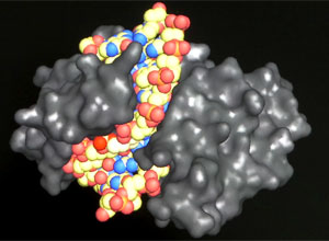
A few years later, in 1970, Hamilton Smith not only independently verified Arber's hypothesis, but also elaborated on the initial discovery by Linn and Arber. He successfully purified a restriction enzyme from another bacterium, Haemophilus influenzae (H. influenzae), and definitively showed that this enzyme cut DNA in the center of a specific six-base-pair sequence. Interestingly, he also showed that this enzyme did not cut at this very same DNA sequence when it occurred in H. influenzae host cell DNA. Building on this result, a first glimpse of how restriction enzymes could be useful tools for scientific research emerged one year later in experiments carried out by Dan Nathans and Kathleen Danna. They used Smith's restriction enzyme to cut the 5,000 base-pair genome of the SV40 virus, which infects monkey and human cells, and identified eleven differently sized pieces of DNA. Nathans's lab later showed that when the SV40 genome was digested with different combinations of restriction enzymes, the sizes of the resulting pieces of DNA could be used to deduce a physical map of the SV40 viral genome, a groundbreaking method for inferring gene sequence information. This method of cutting a DNA molecule into smaller pieces is called a restriction enzyme digest, and it quickly became a powerful tool for generating physical maps of a multitude of genomes, which at the time was a precious revelation in the early stages of genome sequencing. For this groundbreaking set of discoveries, Arber, Smith, and Nathans were jointly awarded the Nobel Prize in Physiology or Medicine in 1978.
Given the vast genetic diversity among bacteria, it follows that different bacterial strains express different restriction enzymes, allowing them to balance their own genes against those of invading bacteriophages. The known variety of restriction enzymes is staggering: To date, more than 4,000 different restriction enzymes that collectively recognize more than 360 different recognition sequences have been isolated from a wide variety of bacterial strains. Based on DNA sequence analysis, scientists know that there are many more restriction enzymes out there waiting to be characterized. The recognition sequences of these enzymes are typically four to six base pairs in length, and they are usually palindromic, which means that their recognition sequence reads the same in the 5' to 3' direction on both DNA strands. There are four different categories of restriction enzymes. Type I restriction enzymes cut DNA at random locations far from their recognition sequence, type II cut within or close to their recognition sequence, type III cut outside of their recognition sequence, and type IV typically recognize a modified recognition sequence.
Type II restriction enzymes, which cut within their recognition sequence, are the most useful for laboratory experiments. Scientists use them to cut DNA molecules at interesting specific locations and then reattach different DNA sequences to each other using an enzyme called DNA ligase, creating new, recombined DNA sequences, or essentially new DNA molecules. This powerful approach to cutting and pasting DNA molecules is known as DNA cloning or recombinant DNA technology. When they act on a DNA molecule, restriction enzymes produce "blunt" ends when they cut in the middle of the recognition sequence, and they yield "sticky" ends when they cut at the recognition sequence in a staggered manner, leaving a 5' or 3' single-stranded DNA overhang. Any two blunt ends can be joined together, but only sticky ends with complementary overhangs can be connected to each other. Restriction enzyme digestion continues to be one of the most common techniques used by researchers who carry out DNA cloning experiments.
Today, researchers rely on restriction enzymes to perform virtually any process that involves manipulating, analyzing, and creating new combinations of DNA sequences. Among the many new combinations are DNA cloning, hereditary disease diagnosis, paternity testing, forensics, genomics (e.g., the human genome project), epigenetics, genetically modified organisms, and biotechnology. Indeed, without the discovery of restriction enzymes, the fields of recombinant DNA technology, biotechnology, and genomics as we know them today would not exist. In 2010, forty years after he purified the first restriction enzyme, Smith was part of the research team that used these very enzymes to build the first synthetic bacterial cell. Led by Craig Venter, this team of scientists used machines to chemically synthesize the one million base-pair Mycoplasma mycoides (M. mycoides) bacterial genome in 1,080 base-pair pieces that were then joined together to form a complete synthetic genome. Along the way, Venter and his colleagues used restriction enzymes to help clone and analyze the synthetic genome. In the final step, they transplanted the synthetic M. mycoides genome into a Mycoplasma capricolum bacterial cell and showed that recipient cells harboring only the synthetic M. mycoides genome were capable of reproducing and exhibited characteristics of M. mycoides cells. In this Spotlight, you'll find a broad range of resources to help you gain a deeper understanding of how restriction enzymes affected the field of molecular biology and our ability to manipulate DNA, as well as how they continue to serve as an invaluable tool for research scientists.
--Heidi Chial, Ph.D. (BioMed Bridge, LLC)
Watch scientists answer questions about the fundamentals of these fascinating enzymes.
Watch an animation that shows how REs “cleave” their DNA targets.

Read about the discovery of REs and how scientists use them.
Learn about how REs are paired with methylases to protect bacterial host DNA while cutting up invading DNA.
Read about how REs operate at the molecular level and how they interact with DNA at the structural level.
Learn about the three major classes of REs, and how the same DNA sequence can be cut differently by specific REs.

Read about how biotechnology relies on REs to recombine DNA fragments and clone genes.
Learn how REs are used for hereditary disease diagnosis, paternity testing, and forensics.
Watch a video about how REs helped sequence the human genome.
Read about how methylation-sensitive REs can be used to identify epigenetic changes in DNA.

Learn how REs play an important role in creating genetically modified organisms.
Read about how REs helped build a synthetic bacterial cell.
Since 1975, this database has organized information about REs, methylases, and the bacteria they originated from.
Richard Roberts, Nobel laureate and founder of REBASE, discusses why it is important to centralize and share RE information.
Watch Hamilton Smith, Nobel laureate for his seminal RE research, discuss the future of synthetic genomes with a student.