This page has been archived and is no longer updated

 
Aa Aa Aa

Scientists Can Make Copies of a Gene through PCR

A schematic shows over 20 double-stranded segments of DNA floating against a pink background. Each DNA segment is composed of two grey, segmented, sugar-phosphate chains oriented opposite from and in parallel to one another. Each segment in a chain represents a single deoxyribose sugar molecule. Nitrogenous bases hang down from the sugar molecules and look like vertical, colored bars. The bars are blue, red, green, or orange. Different colors represent the chemical identity of each nitrogenous base. The bases attached to a strand of DNA nearly meet the ends of the nitrogenous bases on the opposite strand.
Once scientists have zeroed in on a specific segment of DNA, how do they produce enough copies of that segment for their research? In most cases, the polymerase chain reaction, or PCR, is their method of choice for quickly generating a sufficient amount of identical genetic material for study and analysis.

Prior to the development of PCR in the 1980s, the primary method for producing many copies of a gene was a relatively time-consuming process known as DNA cloning. This technique involved insertion of the gene of interest into living bacterial cells, which in turn replicated the gene along with their own DNA during the division and replication processes.

What is PCR?

The key element of PCR is heat. Throughout the PCR process, DNA is subjected to repeated heating and cooling cycles during which important chemical reactions occur. During these thermal cycles, DNA primers bind to the target DNA sequence, enabling DNA polymerases to assemble copies of the target sequence in large quantities.

PCR makes it possible to produce millions of copies of a DNA sequence in a test tube in just a few hours, even with a very small initial amount of DNA. Since its introduction, PCR has revolutionized molecular biology, and it has become an essential tool for biologists, physicians, and anyone else who works with DNA.

How does PCR work?

A schematic illustration shows the equipment needed to perform PCR, including a gray PCR machine, a clear agarose gel electrophoresis chamber connected to a blue and gray power source, and an open microcentrifuge tube half-filled with a dark blue solution. The molecules involved in the reaction are shown, at a disproportionately large size, floating above the equipment. The molecules include a double helix DNA strand; four free-floating nucleotides colored blue, orange, green, and red; and two DNA primers made of 10 nucleotides each.
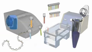
Figure 1: The various components required for PCR include a DNA sample, DNA primers, free nucleotides called ddNTPs, and DNA polymerase.

PCR relies on several key chemical components (Figure 1):

  • A small amount of DNA that serves as the initial template or target sequence
  • A pair of primers designed to bind to each end of the target sequence
  • A DNA polymerase
  • Four dNTPs (i.e., dATP, dCTP, dGTP, dTTP)
  • A few essential ions and salts

The PCR process then uses these ingredients to mimic the natural DNA replication process that occurs in cells. To automate this process, a machine called a thermocycler jump-starts each stage of the reaction by raising and lowering the temperature of the chemical components at specific times and for a preset number of cycles.

Each cycle of PCR has three main steps, as described in the following sections.

Step 1: Denaturation

A schematic shows two rows of nucleotides arranged to form a double-stranded segment of DNA, with half of the nucleotides in the top strand and half of the nucleotides in the bottom strand. Grey horizontal cylinders represent deoxyribose sugar molecules, and blue, red, green, and orange vertical rectangles represent the chemical identity of each nitrogenous base. In the left half of the double-stranded chain, the nitrogenous bases in the upper strand point downward from the sugar-phosphate chain, nearly meeting the ends of the nitrogenous bases from the lower strand. Near the halfway point on the molecule, the two strands appear to be pulling apart and separating, and a larger gap forms between complementary nitrogenous bases on opposing strands.
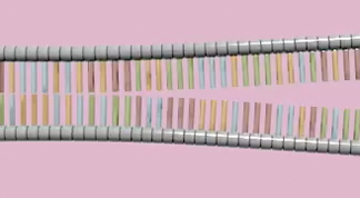
Figure 2: When heated, the DNA strands separate.
During the first step in PCR, the starting solution is heated to the necessary temperature, usually between 90° and 100°C. As the heat builds, it breaks the bonds joining the two strands of the DNA double helix, thereby enabling the DNA to separate into two single strands. This "melting" of the DNA into single strands is called denaturation (Figure 2).

Step 2: Annealing

A schematic shows many nucleotides arranged side-by-side to form a strand of DNA. In each strand, grey horizontal cylinders represent deoxyribose sugar molecules, and blue, red, green, and orange vertical rectangles represent the chemical identity of each nitrogenous base. A second strand of DNA is arranged far below the first strand. The nitrogenous bases of the second strand are facing upward, while the nitrogenous bases of the first strand are facing downward. The two strands of nucleotides represent a double-stranded DNA strand that has been heated to separate the strands and prepare for replication. A 10-nucleotide long primer sequence is shown annealing to the upper strand, and a second primer is shown annealing to the lower strand.
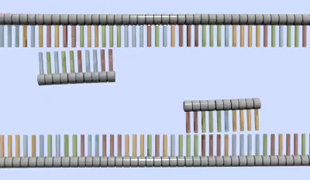
Figure 3: When the solution is cooled, the primers anneal.
After it is held for several minutes at the initial target temperature, the reaction mixture is quickly cooled, usually to between 30° and 65°C. The mixture is then held for less than one minute at this temperature. This gives the primers an opportunity to bind, or anneal, to their complementary sequences on the single strands of DNA (Figure 3).

Step 3: Extension

A schematic shows two DNA molecules: one in the upper half of the frame, and one in the lower half of the frame.  Each DNA molecule is partly single-stranded and partly double-stranded. A transparent blue globular structure, representing the enzyme DNA polymerase, is bound to a several nucleotide-long region along each DNA strand about a quarter of the way from one terminus. The DNA is single-stranded on one side of DNA polymerase and double-stranded on the other side, indicating that DNA polymerase is moving from one terminus towards the other as it builds a new DNA strand from a template sequence. The sugar-phosphate backbone is depicted as a segmented grey cylinder. Nitrogenous bases are represented by blue, orange, red or green vertical rectangles attached above or below each segment of the sugar-phosphate backbone. The nitrogenous bases on one strand bind complementary nucleotides as they are added to the new DNA strand by DNA polymerase. The binding results in red-green or blue-orange pairs of nucleotides, which look like ladder rungs between the grey cylinders.
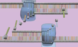
Figure 4: DNA polymerase attaches to each primer and assembles dNTPs to build a new strand.
During the final, or extension, stage of PCR, the sample is heated again, usually to between 60°and 75°C, and it is held at that temperature for less than one minute. At this point, the DNA polymerase begins making a new DNA strand by attaching to the primers and then adding dNTPs to the template strand, thereby creating a complementary copy of the target sequence (Figure 4).

A schematic shows four DNA molecules that have been separated to create eight single-stranded DNA molecules. Each of the single-stranded DNA molecules are undergoing DNA replication.  Each DNA is bound by a transparent blue globular structure, representing the enzyme DNA polymerase. The sugar-phosphate backbone of each strand is depicted as a dark line. Nitrogenous bases are represented by vertical lines attached above or below each segment of the sugar-phosphate backbone. The schematic shows the DNA molecules from a distance, so details in the image are not resolved. Several free-floating replicated DNA strands have been released from their template strand and are awaiting further replication.
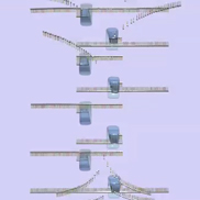
Figure 5: The replication cycle repeats many times.
The number of new copies of the DNA sequence of interest doubles with each three-step cycle. Thus, if the PCR process is repeated 40 or 50 times, even small samples of template DNA can yield millions of identical copies (Figure 5).

PCR is an incredibly versatile technique with many practical applications. Once PCR cycling is complete, the copied DNA molecules can be used for cloning, sequencing, mapping mutations, or studying gene expression.

Variations on conventional PCR

Recently, PCR has proven useful in ways beyond merely copying and propagating identical segments of DNA. Today, geneticists rely on PCR to aid in the study of genes themselves.

Copying and quantifying DNA at the same time using real-time PCR

One modification of conventional PCR allows researchers to copy a particular DNA sequence and quantify it simultaneously. Dubbed quantitative real-time PCR (qPCR), this technique makes it possible to measure the amount of DNA produced during each PCR cycle. This refinement involves the use of fluorescent dyes or probes that label double-stranded DNA molecules. These fluorescent markers bind to the new DNA copies as they accumulate, making "real-time" monitoring of DNA production possible.

As the number of gene copies increases with each PCR cycle, the fluorescent signal becomes more intense. Plotting fluorescence against cycle number and comparing the results to a standard curve (produced by real-time PCR of known amounts of DNA) enables scientists to determine the amount of DNA present during each step of the PCR reaction.

Quantifying RNA using reverse transcription PCR

Real-time PCR can also be used to calculate the amount of specific kinds of genetic material other than DNA, such as RNA. This extension of real-time PCR technology, called reverse transcription PCR (RT-PCR), combines real-time PCR with reverse transcription, the process that makes DNA from mRNA. RT-PCR can be used to determine how gene expression changes over time or under different conditions. For this reason, this technique is sometimes used to verify microarray data.

Watch this video for a summary of the PCR process


Further Exploration


Connect
Connect Send a message


Scitable by Nature Education Nature Education Home Learn More About Faculty Page Students Page Feedback


Genetics

Visual Browse

Close