


« Prev Next »

Replication and Distribution of DNA during Mitosis

Walther Flemming's drawing of chromosomes.
Most cells grow, perform the activities needed to survive, and divide to create new cells. These basic processes, known collectively as the cell cycle, are repeated throughout the life of a cell. Of the various parts of the cell cycle, the division portion is particularly important, because this is the point at which a cell passes its genetic information to its offspring cells. In many situations, division also ensures that new cells are available to replace the older cells within an organism whenever those cells die.
Prokaryotic cells, which include bacteria, undergo a type of cell division known as binary fission. This process involves replication of the cell's chromosomes, segregation of the copied DNA, and splitting of the parent cell's cytoplasm. The outcome of binary fission is two new cells that are identical to the original cell.
In contrast to prokaryotic cells, eukaryotic cells may divide via either mitosis or meiosis. Of these two processes, mitosis is more common. In fact, whereas only sexually reproducing eukaryotes can engage in meiosis, all eukaryotes — regardless of size or number of cells — can engage in mitosis. But how does this process proceed, and what sorts of cells does it produce?
What happens during mitosis?
During mitosis, a eukaryotic cell undergoes a carefully coordinated nuclear division that results in the formation of two genetically identical daughter cells. Mitosis itself consists of five active steps, or phases: prophase, prometaphase, metaphase, anaphase, and telophase. Before a cell can enter the active phases of mitosis, however, it must go through a period known as interphase, during which it grows and produces the various proteins necessary for division. Then, at a critical point during interphase (called the S phase), the cell duplicates its chromosomes and ensures its systems are ready for cell division. If all conditions are ideal, the cell is now ready to move into the first phase of mitosis.
Prophase

Figure 1: During prophase, the chromosomes in a cell's nucleus condense to the point that they can be viewed using a light microscope.
Prophase is the first phase of
mitosis. During this phase, the chromosomes inside the cell's nucleus condense and
form tight structures. In fact, the chromosomes become so dense that they
appear as curvy, dark lines when viewed under a microscope (Figure 1). Because
each chromosome was duplicated during S phase, it now consists of two identical
copies called sister chromatids that are attached at a common center point called the centromere.
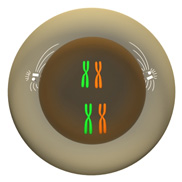
Figure 2: The mitotic spindle (white) begins to form outside the cell's nucleus.
Important changes also take place outside of the nucleus during
prophase. In particular, two structures called centrosomes move to opposite sides of the cell during this phase
and begin building the mitotic spindle.
The mitotic spindle plays a critical role during the later phases of mitosis as it orchestrates the movement of sister chromatids to opposite poles of the cell (Figure 2).
Prometaphase
After prophase is complete, the cell enters prometaphase. During prometaphase, the
nuclear membrane disintegrates and the mitotic spindle gains access to the chromosomes. During this phase, a protein structure called the kinetochore is associated with the centromere on each sister chromatid.
Stringlike structures called microtubules
grow out from the spindle and connect
to the sister chromatids at their kinetochores; one microtubule from one side
of the spindle attaches to one sister chromatid in each chromosome, and one
microtubule from the other side of the spindle attaches to the other sister
chromatid (Figure 3a).
Metaphase
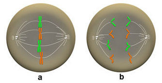
Figure 3: (a) Metaphase and (b) Anaphase.
In metaphase (a), the microtubules of the spindle (white) have attached and the chromosomes have lined up on the metaphase plate. During anaphase (b), the sister chromatids are pulled apart and move toward opposite poles of the cell.
Anaphase
After metaphase is complete, the cell enters anaphase.
During anaphase, the microtubules attached to the kinetochores
contract, which pulls the sister chromatids apart and toward opposite
poles of the cell (Figure 3c). At this point, each chromatid is
considered a separate chromosome.
Telophase
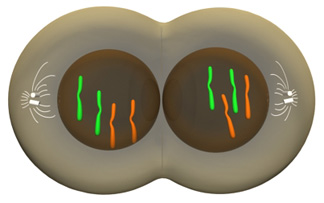
Figure 4: During telophase, two nuclear membranes form around the chromosomes, and the cytoplasm divides.
Finally, once anaphase is complete, the cell enters the last stage of the division process — telophase.
During telophase, the newly separated chromosomes reach the mitotic
spindle and a nuclear membrane forms around each set of chromosomes,
thus creating two separate nuclei inside the same cell. As Figure 4 illustrates, the cytoplasm
then divides to produce two identical cells.
Why is mitosis important?
As previously mentioned, most eukaryotic cells
that are not involved in the production of gametes undergo mitosis. These
cells, known as somatic cells, are
important to the survival of eukaryotic organisms, and it is essential that
somatic parent and daughter cells do not vary from one another. With few exceptions,
the mitotic process ensures that this is the case. Therefore, mitosis ensures that
each successive cellular generation has the same genetic composition as the
previous generation, as well as an identical chromosome set.
Further Exploration
eBooks
This page appears in the following eBook


