


« Prev Next »

Mitochondria
Mitochondria are unusual
organelles. They act as the power plants of the cell, are surrounded by two
membranes, and have their own genome. They also divide independently of the
cell in which they reside, meaning mitochondrial replication is not coupled
to cell division. Some of these features are holdovers from the
ancient ancestors of mitochondria, which were likely free-living prokaryotes.
What Is the Origin of Mitochondria?
Mitochondria are thought to have originated from an ancient symbiosis that resulted when a nucleated cell engulfed an aerobic prokaryote. The engulfed cell came to rely on the protective environment of the host cell, and, conversely, the host cell came to rely on the engulfed prokaryote for energy production. Over time, the descendants of the engulfed prokaryote developed into mitochondria, and the work of these organelles — using oxygen to create energy — became critical to eukaryotic evolution (Figure 1).
Modern mitochondria have striking similarities to some modern prokaryotes, even though they have diverged significantly since the ancient symbiotic event. For example, the inner mitochondrial membrane contains electron transport proteins like the plasma membrane of prokaryotes, and mitochondria also have their own prokaryote-like circular genome. One difference is that these organelles are thought to have lost most of the genes once carried by their prokaryotic ancestor. Although present-day mitochondria do synthesize a few of their own proteins, the vast majority of the proteins they require are now encoded in the nuclear genome.
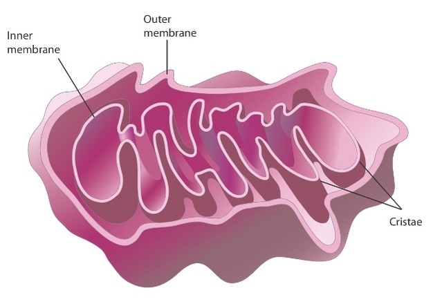
What Is the Purpose of a Mitochondrial Membranes?
As previously mentioned, mitochondria contain two major membranes. The outer mitochondrial membrane fully surrounds the inner membrane, with a small intermembrane space in between. The outer membrane has many protein-based pores that are big enough to allow the passage of ions and molecules as large as a small protein. In contrast, the inner membrane has much more restricted permeability, much like the plasma membrane of a cell. The inner membrane is also loaded with proteins involved in electron transport and ATP synthesis. This membrane surrounds the mitochondrial matrix, where the citric acid cycle produces the electrons that travel from one protein complex to the next in the inner membrane. At the end of this electron transport chain, the final electron acceptor is oxygen, and this ultimately forms water (H20). At the same time, the electron transport chain produces ATP. (This is why the the process is called oxidative phosphorylation.)
During electron transport, the participating protein complexes push protons from the matrix out to the intermembrane space. This creates a concentration gradient of protons that another protein complex, called ATP synthase, uses to power synthesis of the energy carrier molecule ATP (Figure 2).
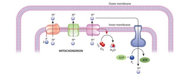
Figure 2: The electrochemical proton gradient and ATP synthase
At the inner mitochondrial membrane, a high energy electron is passed along an electron transport chain. The energy released pumps hydrogen out of the matrix space. The gradient created by this drives hydrogen back through the membrane, through ATP synthase. As this happens, the enzymatic activity of ATP synthase synthesiszes ATP from ADP.
© 2010 Nature Education All rights reserved. 
Is the Mitochondrial Genome Still Functional?
Mitochondrial genomes are very small and show a great deal of variation as a result of divergent evolution. Mitochondrial genes that have been conserved across evolution include rRNA genes, tRNA genes, and a small number of genes that encode proteins involved in electron transport and ATP synthesis. The mitochondrial genome retains similarity to its prokaryotic ancestor, as does some of the machinery mitochondria use to synthesize proteins. In fact, mitochondrial rRNAs more closely resemble bacterial rRNAs than the eukaryotic rRNAs found in cell cytoplasm. In addition, some of the codons that mitochondria use to specify amino acids differ from the standard eukaryotic codons.
Still, the vast majority of mitochondrial proteins are synthesized from nuclear genes and transported into the mitochondria. These include the enzymes required for the citric acid cycle, the proteins involved in DNA replication and transcription, and ribosomal proteins. The protein complexes of the respiratory chain are a mixture of proteins encoded by mitochondrial genes and proteins encoded by nuclear genes. Proteins in both the outer and inner mitochondrial membranes help transport newly synthesized, unfolded proteins from the cytoplasm into the matrix, where folding ensues (Figure 3).
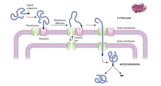
Figure 3: Protein import into a mitochondrion
A signal sequence at the tip of a protein (blue) recognizes a receptor protein (pink) on the outer mitochondrial membrane and sticks to it. This causes diffusion of the tethered protein and its receptor through the membrane to a contact site, where translocator proteins line up (green). When at this contact site, the receptor protein hands off the tethered protein to the translocator protein, which then channels the unfolded protein past both the inner and outer mitochondrial membranes.
© 2010 Nature Education All rights reserved.
How Many Mitochondria Do Cells Have?
Mitochondria cannot be made "from scratch" because they need both
mitochondrial and nuclear gene products. These organelles replicate by
dividing in two, using a process similar to the simple, asexual form of
cell division employed by bacteria. Video microscopy shows that
mitochondria are incredibly dynamic. They are constantly dividing,
fusing, and changing shape. Indeed, a single mitochondrion may contain
multiple copies of its genome at any given time.
Conclusion
Mitochondria, the so-called "powerhouses" of cells, are unusual
organelles in that they are surrounded by a double membrane and retain
their own small genome. They also divide independently of the cell cycle
by simple fission. Mitochondrial division is stimulated by
energy demand, so cells with an increased need for energy contain
greater numbers of these organelles than cells with lower energy needs.


