


« Prev Next »

Ion Channel
Certain cells, commonly called excitable cells, are unique because of their ability to generate electrical signals. Although several types of excitable cells exist — including neurons, muscle cells, and touch receptor cells — all of them use ion channel receptors to convert chemical or mechanical messages into electrical signals.
Like all cells, an excitable cell maintains a different concentration of ions in its cytoplasm than exists in its extracellular environment. Together, these concentration differences create a small electrical potential across the plasma membrane. Then, when conditions are right, specialized channels in the plasma membrane open and allow rapid ion movement into or out of the cell, and this movement creates an electrical signal. But what do these channels look like, and how do they function? Also, how do the electrical signals generated by excitable cells differ from the other types of signals involved in cellular communication?
What Are Ion Channel Receptors?
Ion channel receptors are usually multimeric proteins located in the plasma membrane. Each of these proteins arranges itself so that it forms a passageway or pore extending from one side of the membrane to the other. These passageways, or ion channels, have the ability to open and close in response to chemical or mechanical signals. When an ion channel is open, ions move into or out of the cell in single-file fashion. Individual ion channels are specific to particular ions, meaning that they usually allow only a single type of ion to pass through them. Both the amino acids that line a channel and the physical width of the channel determine which ions are able to wiggle through from the cell exterior to its interior, and vice versa. The opening of an ion channel is a fleeting event. Within a few milliseconds of opening, most ion channels close and enter a resting state, where they are unresponsive to signals for a short period of time (Figure 1).
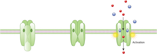
Figure 1: An example of ion channel receptor activation
An acetylcholine receptor (green) forms a gated ion channel in the plasma membrane. This receptor is a membrane protein with an aqueous pore, meaning it allows soluble materials to travel across the plasma membrane when open. When no external signal is present, the pore is closed (center). When acetylcholine molecules (blue) bind to the receptor, this triggers a conformational change that opens the aqueous pore and allows ions (red) to flow into the cell.
© 2010 Nature Education All rights reserved. 
How Are Electrical Signals Propagated?
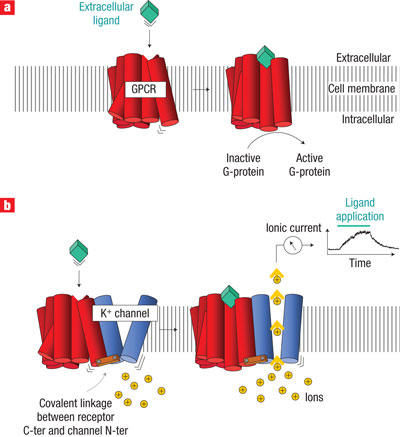
Figure 2: Comparing the activation of an ion channel receptor with that of a G-protein-coupled receptor
Activation of both a G-protein-coupled receptor (a) and an ion channel receptor (b) cause a conformational change in the receptor protein. G protein activation can lead to multiple intracellular events through a variety of intracellular proteins, and this signaling can take seconds to minutes. When a G protein activates transcription, this can take up to 20 minutes. In contrast, ion channel receptors open pores in the cell membrane, causing the formation of electrical current. This receptor activation therefore causes a much faster response within the cell, on the order of milliseconds.
© 2008 Nature Publishing Group Moreau, C. J. et al. Coupling ion channels to receptors for biomolecule sensing. Nature Nanotechnology 3, 620-625 (2008) doi:10.1038/nnano.2008.242. All rights reserved.
Electrical signals travel much more rapidly than chemical signals, which depend on the process of molecular diffusion. As a consequence, excitable cells respond to signals much more rapidly than cells that rely solely on chemical signals (Figure 2). In fact, an electrical signal can traverse the entire length of a human nerve cell — a distance of as much as one meter — within only milliseconds.
How Do Different Types of Excitable Cells Work?
Neurons, muscle cells, and touch receptor cells are all excitable cells — which means they all have the capacity to transmit electrical signals. Each of these cells also has ion channel receptors clustered on a particular part of its surface. For example, the receptors that respond to chemical signals are generally located at synapses — or points of near contact between adjacent cells.
Of the various types of excitable cells that respond to chemical signals, neurons are perhaps the most familiar. When electrical signals reach the end of neurons, they trigger the release of chemical messengers called neurotransmitters. Each neurotransmitter then diffuses from its point of release on one side of the synapse to the cell on the other side of the synapse. If the neurotransmitter binds to an ion channel receptor on the target cell, the related ion channel opens, and an electrical signal propagates itself along the length of the target cell.
Neurons have ion channel receptors specific to many kinds of neurotransmitters. Some of these neurotransmitters act in an excitatory capacity, bringing their target cells ever closer to signal propagation. Other neurotransmitters exert an inhibitory effect, counteracting any excitatory input and lessening the chance that the target cell will fire.
Skeletal muscle cells also rely on chemical signals in order to generate electrical signals. These cells have synapses that are packed with receptors for acetylcholine, which is the primary neurotransmitter released by motor neurons. When acetylcholine binds to the receptors on a skeletal muscle cell, ion channels in that cell open, and this launches a sequence of events that results in contraction of the cell.
In contrast to neurons and skeletal muscle cells, some excitable cells have ion channels that open in response to mechanical stimuli rather than chemical signals. These include the hair cells of the mammalian inner ear and the touch receptor cells of both human finger pads and Venus fly traps. Cells that respond to touch have their ion channel receptors clustered at the position where contact usually occurs.
Conclusion
Excitable
cells, such as fast-acting neurons and muscle cells, have specialized
channels
that open in response to a signal and permit rapid ion movement across
the cell
membrane. The opening of just a single ion channel alters the electrical
charge
on both sides of the membrane. The resulting charge differential then
causes
adjacent voltage-sensitive channels to open in chain-reaction fashion,
creating
a self-propagating electrical signal that travels down the entire length
of the
cell. Sometimes, this sequence of events is triggered when a chemical
signal — such as a neurotransmitter — binds to an ion channel receptor
on cell's
surface. Other times, a cell's ion channels open in response to
mechanical
(rather than chemical) stimuli.
eBooks
This page appears in the following eBook


