


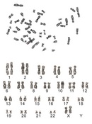
 Figure 1


« Prev Next »
The rediscovery of Mendel's laws near the beginning of the twentieth century triggered intense interest in the principles of heredity. The chromosome theory of heredity was proven early in the century; meanwhile, a great deal of scientific interest was directed toward learning more about chromosomes themselves. However, defining the human diploid chromosome number would prove more challenging than investigators initially anticipated.
Difficulties in determining the human diploid number arose for a variety of reasons. For one, early experiments that provided evidence for the chromosome theory often used invertebrate species that reproduced in large numbers and had a relatively low number of well-defined chromosomes. Neither of these characteristics, of course, is a common finding in humans. In addition, the human samples initially used for chromosome analysis were derived from fresh testicular tissue in which haploid meiotic cells were often present. Furthermore, what morphology could be deduced suggested that human chromosomes were more complex than those of the model organisms studied earlier. In light of these and other factors, an erroneous estimate by prominent cytologist Theophilus Painter dominated the field for decades, until researchers Joe Hin Tjio and Albert Levan eventually applied new technology to identify the true diploid number of human chromosomes.
Theophilus Painter Reports that Humans Have 48 Chromosomes
Theophilus Painter was one of the preeminent cytologists of the early twentieth century. Like many cytologists of the time, Painter was highly interested in human heredity, and this interest fueled his attempts to determine the diploid number of human chromosomes.
Painter's Approach
Painter began his investigation of human chromosome number by obtaining samples of human testicular tissue, which were embedded in paraffin and then sliced into thin sections. Next, he transferred these serial sections to glass microscope slides and stained them to allow visualization of the chromosomes. The very nature of these experiments meant that it was rare for all chromosomes in a given nucleus to be visualized simultaneously. As a result, reconstruction of intact nuclei was necessary, and it involved assembly of data from consecutive sections. To further complicate matters, the tissues studied by Painter were obtained from a single institutionalized individual who was likely to have had constitutional numerical chromosome aberrations. As such, Painter's report of a human diploid chromosome number of 48 in 1923 had more than one possible source of error.
Analysis of Painter's Methods
By looking at Painter's drawings of his slides, one can appreciate how difficult this process made it to arrive at a correct chromosome count. For example, Frank Ruddle (2004), a well-respected modern cytologist, speculates that Painter failed to identify human chromosome 1 as a single chromosome because of a staining artifact. Chromosome 1 is a large, metacentric chromosome with a considerable amount of heterochromatin at its centromere. Ruddle speculates that this heterochromatin failed to take up the iron hematoxylin stain that Painter was using. Consequently, the heterochromatin appeared as a gap between two chromosomes. Supporting this argument, Ruddle notes that chromosome 1 appears to be missing from Painter's ordered display of chromosomes in his 1923 paper. Arrows that Ruddle added to Painter's original figure point to "chromosomes" that may actually be the two arms of chromosome 1, with centromeres lacking and the two arms approximating the right sizes for chromosome 1. This error notwithstanding, Painter's estimate was very close to the real human diploid number of 46, and the quality of his data was good. In light of Painter's many other contributions to cytology, the scientific community accepted his estimate of the human chromosome number for 33 years.
Tjio and Levan Use Improved Methods to Establish the Chromosome Number as 46
In the decades following Painter's work, scientists continued to refine their methods for preparing chromosomes for microscopy. Sectioning of paraffin-embedded preserved tissue was gradually replaced by squash techniques, in which small tissue specimens are placed on a microscope slide and then literally squashed under a cover slip to produce a single layer of cells. This approach gained broad acceptance as it eliminated any need to slice through tissues and reconstruct the organization of chromosomes in a single nucleus from several different sections. Chromosome preparations were also dramatically improved by combining treatment with a hypotonic salt solution (described by T. C. Hsu in 1952) and cell fixation. This combination of treatments enhanced chromosome spreading without deterioration or fragmentation, thereby facilitating better chromosome counts. In fact, in 1956, these techniques enabled researchers Joe Hin Tjio and Albert Levan to make a more accurate estimate of the human chromosome number.
Tjio and Levan’s Approach
When their classic paper was published in 1956, Tjio and Levan had already been collaborating for several years. Albert Levan was a well-established cytologist who had pioneered the use of colchicine for analyzing chromosomes. Colchicine is a plant-derived toxin that arrests cells in metaphase, the point in the cell cycle at which chromosomes are most condensed. Colchicine is toxic to animals, but Levan and others found that colchicine allowed investigators to work with cells grown in tissue culture. Capturing cells at a specific state of mitosis when the chromosomes are condensed and easily tracked improved the reliability of their observations. A sample metaphase chromosome spread produced using this method is shown in Figure 1.
Tjio and Levan used spreads such as these in their research, eventually reporting summary data from 261 unique chromosome spreads obtained from 22 different cell cultures of fetal lung tissue. All of the cultures were used within a few days after the tissue was obtained, thus minimizing the possibility of long-term culture-induced artifacts of chromosome number. The results were both clear and replicable. In the words of Tjio and Levan, "We were surprised to find that the chromosome number 46 predominated in the tissue cultures from all four embryos, [with] only single cases deviating from this number." Appreciating the fact that these in vitro data may not have been representative of cells in the body (i.e., in vivo data), Tjio and Levan also highlighted the importance of finding the same chromosome number in spermatogenic cells from testicular samples. Within a year, Ford and Hamerton (1956) did just that, providing confirmatory data by reporting the diploid chromosome number in human testicular cells to be 46.
Analysis of Tjio and Levan’s Methods
By today's standards, Tjio and Levan's initial chromosome preparations offered relatively poor resolution of metaphase chromosomes. The gross morphology of the chromosomes was apparent, but few other distinguishing features were clear. Nonetheless, Tjio and Levan's determination of a human diploid number of 46 chromosomes was proven correct.
Over the next several decades, better technology made it possible to both confirm and expand upon Tjio and Levan's results. For instance, a variety of banding techniques that were introduced during the 1970s offered increased resolution and allowed individual chromosomes to be distinguished from one another. Today, banding techniques such as Giemsa-trypsin based staining are commonly used in diagnostic cytogenetics, and they can provide a resolution greater than 5 Mb. In addition, more sophisticated (and sometimes targeted) molecular cytogenetic analyses now offer even greater resolution for diagnostic purposes (Trask, 2002).
References and Recommended Reading
Ford, C. E., & Hamerton, J. L. The chromosomes of man. Nature 178, 1020–1023 (1956) doi:10.1038/1781020a0 (link to article)
Gartler, S. M. The chromosome number in humans: A brief history. Nature Reviews Genetics 7, 655–660 (2006) doi:10.1038/nrg1917 (link to article)
Hsu, T. C. Mammalian chromosomes in vitro, I. Karyotype of man. Journal of Heredity 43, 167–172 (1952)
Painter, T. S. Studies in mammalian spermatogenesis II: The spermatogenesis of man. Journal of Experimental Zoology 37, 291–336 (1923)
Ruddle, F. H. Theophilus Painter: First steps toward an understanding of the human genome. Journal of Experimental Zoology 301A, 375–377 (2004) doi:10.1002/jez.a.20072
Tjio, J. H., & Levan, A. The chromosome number of man, Hereditas 42, 1–6 (1956)
Trask, B. J. Human cytogenetics: 46 chromosomes, 46 years and counting. Nature Reviews Genetics 3, 769–778 (2002) doi:10.1038/nrg905 (link to article)


