Science Photography


On January 7, 1839, an installation artist and chemist named Louis-Jacques-Mandé Daguerre announced to the French Academy of Sciences in Paris that he had perfected a photographic imaging technology that could be used on a large scale and was reproducible. His invention, later named the daguerreotype, was based on a special property of silver iodide: when exposed to light, the molecules undergo a chemical transformation that can be turned into charcoal-colored shadows and lines when later exposed to developing mercury fumes. The more intense the light, the darker the resulting silver-mercury amalgam. When a sheet of metal copper coated with silver iodide, for instance, was exposed to a street scene for a prolonged period, an image of the street was represented on the daguerreotype plate as lines and shadows of varying brightness.
Daguerre initially thought his invention would be suitable mostly for personal use in travel logs, art installations, and architectural records. But it was increasingly used in scientific observation throughout the nineteenth century, due to its ability to capture natural reality in a single moment and preserve it for extended scrutiny. Astronomers were among the first to embrace this technique; the first images of the sun and of a solar eclipse were both captured on daguerreotypes in the 1840s. By the end of the nineteenth century, photographic images — by then based on a newer technique of transferring images from silver-coated glass plates to paper — had become a core tool of the scientific trade, and photographic images were appearing frequently in scientific journals. Since then, the role of photography in science has only grown.
Why have scientists always regarded photography so highly? Careful observation of evidence is the heart of modern scientific method; photography has always been valued as an objective technique of observation, freed from the potential for human error implicit in the older method of sketching experimental observations. Just as important, photography can gather data that can't be detected or processed by the human eye. Using technology that captured the scattered path of invisible x-ray beams, for example, Rosalind Franklin in 1952 was able to reveal the precise structure of intertwined DNA molecules — what we now recognize as the double helix. At the other end of the spatial scale, telescopic cameras can record galaxies located four billion light years away from Earth, as in these images from NASA's Chandra X-ray Observatory. And photography can even "slow down" events that are ordinarily too quick for the eye. In 1878, for example, Eadweard Muybridge's revolutionary photographs of a horse in motion settled a longstanding dispute about whether all four of a running horse's feet are ever off the ground at the same time (they are). In the 1950s, MIT physicist Harold Edgerton used a strobe light flash to produce stunningly beautiful images of the precise patterns made by a bullet passing through an apple, a drop of milk splashing into a glass, and a hummingbird flapping its wings.
These discoveries illustrate a fundamental pattern: technological advances in optics, cameras, and the control of lighting enable scientific advance and discovery. A recent example of this dynamic involves digital cameras. Developed in the early 1980s, computerized cameras that use a light-sensitive electronic chip called a charge-coupled device (CCD) allowed wavelengths of light to be converted into electronic charges (rather than into oxidized silver grains) and enabled more sensitive control over exposure times. Scientists quickly realized that, because they can capture images in very dark conditions, CCD cameras could be used to record shapes and structures deep inside biological tissue or under a microscope lens.
Today scientists and engineers continue to work together to develop more powerful scientific imaging technologies, such as functional magnetic resonance imaging (fMRI), which measures the magnetic fields of oxygen molecules deep inside brain tissue in order to track brain activity, and atomic force microscopy (AFM), which uses reflected laser beams to map the tiny, intricate surfaces of linked carbon atoms in nanomaterials. The stories of science and of photography are certain to be intertwined throughout the twenty-first century. In this Spotlight, we invite you to learn about the forms of photography that contribute to cutting-edge scientific advancement.
What is Science Photography?
This interactive timeline shows important moments in photography's history.
Listen to curator Corey Keller describe how the 1800s changed photography.
Read how our ability to image the stars allowed a new science to emerge.
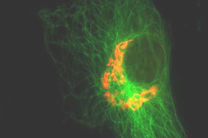
Microscope Photography
Read this introduction to photomicrography provided by the Optical Microscopy Division of the National High Field Laboratory, a joint venture of FSU, UF, and the Los Alamos National Laboratory.
The Institute for Molecular Virology at the University of Wisconsin at Madison provides a gallery of images of viruses, bacteriophages, DNA, and DNA-protein complexes as well as its methods.
Listen to scientist Stephen J. Pennycock explain how microscopy can help us see the ways atoms are bonded together in this 2004 interview with NPR.
Learn about Hubble, named in honor of astronomer Edwin Hubble, which was launched in 1990 and captures both visible and some UV light.
Used during the 1990s, CGRO captured "hard" x-rays emitted by the nucleus of an atom.
Since its launch in 1999, the Chandra X-ray Observatory has been NASA's flagship mission for x-ray astronomy.
During its mission begun in 2003, Spitzer obtains images and spectra by detecting the infrared energy, or heat, radiated by objects in space.
Launching in 2014, the James Webb Space Telescope will detect infrared radiation and be capable of seeing in that wavelength as well as Hubble sees in visible light.
Created through a partnership between NASA and the Internet Archive, NASA Images offers public access to the largest source of NASA's image, video, and audio collections.
Opportunity and Education in Science Photography
The National Science Foundation and the journal Science created the challenge to celebrate that grand tradition — and to encourage its continued growth.
Learn how for more than thirty years, Nikon has rewarded the world's best photomicrographers who make critically important scientific contributions to life science, bioresearch, and materials science.
Dennis Kunkel Microscopy, Inc. is an education resource that provides background information, an image library, and a virtual Scanning Electron Microscope (SEM).
, and T.A. Rector (NRAO)")


