Volume 9
-
No. 12 December 2002
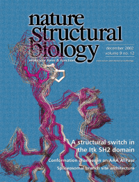
Overlay of the solution structures of the interleukin-2 tyrosine kinase (Itk) SH2 domain. A single proline imide bond (Pro is highlighted as a ball-and-stick model) in this domain adopts both cis and trans conformations, creating structures (white, cis; red, trans) that have different substrate recognition specificities. Figure created by A. Laederach. See pages 900–905.
-
No. 11 November 2002
The structure of the penicillin binding protein PBP2a (ribbon and surface) from methicillin-resistant Staphylo-coccus aureus (MSRA). The location of the active site in the blue transpeptidase domain is indicated by the red nitrocefin adduct (stick rendering). PBP2a confers broad spectrum resistance to all clinically used β-lactam antibiotics. The protein surface image was prepared by G. Olovsson; S. Farmer, C. Friedrich and R. Hancock provided the scanning elecron micrograph of MSRA (background). See pages 870–876.
-
No. 10 October 2002
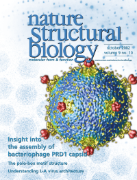
Mobile arms and glue proteins hold together the capsid of PRD1, a broad host-range bacteriophage. The foreground is the difference image between the cryo-EM reconstruction of the phage and a quasi-atomic model of the capsid derived from the structure of the major coat protein (P3). The vertex complexes are red, flexible P3 arms and glue proteins are yellow, and the viral membrane is blue. The background symbolizes the merging of the two techniques, the X-ray diffraction pattern of P3 crystals (top) and a typical PRD1 cryo-electron micrograph (bottom). X-ray diffraction image courtesy of S.D. Benson. See pages 756–763.
-
No. 9 September 2002
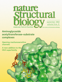
Mycobacterium tuberculosis amino-glycoside 2'-N-acetyltransferase (green surface and ribbons) in complex with cofactor coenzyme A (red) and antibiotic substrate ribostamycin (yellow). This enzyme can modify a broad range of aminoglycoside drugs and thereby confer antibiotic resistance to the bacteria. The structure provides insights into the modification reaction mechanism. It also suggests that the enzyme may participate in regulating the redox potential in mycobacterial cells. See pages 653–658.
-
No. 8 August 2002
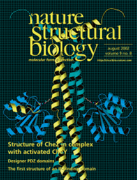
Crystal structure of CheZ (yellow and green) in complex with its substrate, activated CheY (blue; the phosphate mimic, BeF 3- , is shown as a yellow and purple ball-and-stick model). CheZ is the last component in the Escherichia coli chemotaxis pathway without a high resolution structure. The structure of the complex provides insights into how CheZ enhances dephosphorylation of CheY-phosphate. See pages 570-575, and News and Views pages 563–564.
-
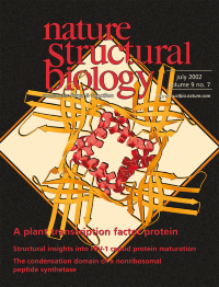
No. 7 July 2002
The structure of the p24 subunit of the plant defense transcription factor PBF-2. p24 is a prototype of a new structural family, the ‘Whirly’ family, so named for the whirligig quaternary structure. The tetrameric architecture of p24 displays a novel single-stranded DNA binding surface and thus may have a new DNA recognition mechanism. See pages 512–517.
-
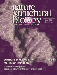
No. 6 June 2002
The structure of the U6 RNA intra-molecular stem loop of S. cerevisiae (stick model with transparent surface) reveals a stereo-specific binding site for a metal ion (modeled, purple ball) implicated in catalysis. The structure provides the first view of U6 RNA, a key component of the spliceosome, and suggests a possible mechanism for the regulation of RNA splicing. Background is from the 2D NOE spectrum of the U6 ISL. See pages 431–435.
-
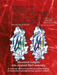
No. 5 May 2002
Mapping of the core structure in ß2-microglobulin amyloidogenic pre-cursor and in the assembled fibril. When mapped onto the native protein, the core regions in the precursor (left; blue) are similar to those in the fibril (right, purple). Together, these studies provide structural insights into the two extremes of the amyloid assembly process. The background is an electron micrograph of ß2-microglobulin fibrils. See pages 326–331 and 332–336; News and Views pages 323–325.
-
No. 4 April 2002
Spotlight on the radical generating apparatus in L. leichmannii class II ribonucleotide reductase (surface map) with bound adeninopentylcobalamin (stick model). The cobalamin analog is located ∼ 10 å from the site of the thiyl radical (pink). The structure provides the first view of the arrangement of all the key factors in radical generation in RNR and insight into the allosteric regulation of a monomeric protein. See pages 293-300 and News and Views pages 236–238.
-
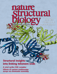
No. 3 March 2002
Structure of the telomere end-binding protein α-subunit (ribbon diagram) in complex with single-stranded DNA (stick model) containing telomeric sequence. The oligonucleotides are at opposite sides of the α-subunit homodimer, suggesting that the protein can simultaneously bind two telomeres and mediate telomere—telomere interactions to form rosette-like structures of macronuclear chromatin (background, electron micrograph courtesy of D. Prescott and G. Murti).
-
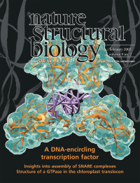
No. 2 February 2002
TonEBP is a transcription factor that regulates gene expression induced by osmotic stress in mammalian cells. The crystal structure of TonEBP (orange ribbon in glass surface) in complex with a target DNA (purple) reveals that the transcription factor binds as a dimer and completely wraps around the DNA. This binding mode retards dissociation of the complex and may be the major mechanism of TonEBP binding to DNA. See pages 90-94.
-
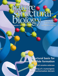
No. 1 January 2002
Shown on the cover is the active site of Erv2p, a sulfhydryl oxidase that promotes disulfide bond formation in the ER. The structure, which differs from other known FAD-binding proteins, reveals the location of two pairs of cysteines (shown in yellow) required for activity. One of these cysteine pairs is in the flexible C-terminal arm of the protein and may shuttle newly generated disulfides to substrate proteins. See pages 61–67 and News and Views pages 2–3