
Volume 26 Issue 12, December 2019
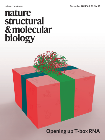
Opening up T-box RNA
Structural elucidation of T-box–tRNA complexes provides new insights into the mechanisms of tRNA decoding and aminoacylation sensing by T-box riboswitches.
See Li, Su et al., Battaglia et al., Suddala and Zhang
Image: composite by Erin Dewalt using image from Yevgen Romanenko / Moment / Getty and structures of T-box–tRNA complex in ribbon representation prepared by Jacob Weaver and Alexander Serganov. Cover Design: Erin Dewalt.
News & Views
-
Advertisement
