Volume 12
-
No. 12 December 2005
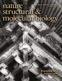
A Japanese Buddhist temple uses an elaborate system of brackets (cover image) in its architecture. Hamma et al. show that Cbf5 and Nop10, components of the box H/ACA ribonucleoprotein complex involved in pseudouridine modification, have a conserved architecture that may function to organize the secondary structure of the H/ACA RNAs. Photo by R. Reeder. pp 1101-1107
-
No. 11 November 2005
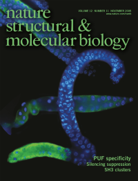
Stem cells proliferate at one end of the C. elegans germline, and sperm differentiate at the other. FBF, a pumilio-like protein, promotes proliferation in the germline by repressing gld-1 mRNA. Opperman et al. show that FBF requires a single 'extra' nucleotide in its RNA targets, and so specifically binds gld-1 and not other RNAs. Blue is DAPI, green is a sperm marker. Images provided by S. Crittenden and J. Kimble. pp 945-951
-
No. 10 October 2005
Sec15 is a component of the exocyst complex involved in vesicle transport and localization. Sec15 recognizes vesicles associated with Rab GTPases. Quiocho and colleagues show that the C-terminal helical domain of Drosophila Sec15 interacts with Rab11 in a GTP-dependent manner, co-localizes with this Rab GTPase in specialized structures within the fly eye called rhabdomeres (cover), and is important for rhabdomere morphology. pp 879-885
-
No. 9 September 2005
Translation elongation factor 1A (eEF1A) has a role in protein synthesis and binds to the actin cytoskeleton. Gross and Kinzy show that these functions are separable and identify specific regions of eEF1A that are important for actin binding in vivo and bundling in vitro. The bamboo trunks shown on the cover resemble the unbundled actin in cells expressing mutant eEF1A. pp 772-778
-
No. 8 August 2005
The red ribbon symbolizes the global solidarity with people living with HIV/AIDS and the commitment to fight this disease. Here, the ribbon puzzle represents the HIV capsid assembly, which is essential for the integrity of the immature and mature virus particle. Individual capsid protein dimers form the puzzle pieces. Binding of a newly identified peptide inhibitor alters the pieces such that they cannot assemble into infectious particles. The peptide is thus a lead compound for a new class of HIV drugs. pp 671-677, pp 678-682; News and Views pp 638-639.
-
No. 7 July 2005
Conus snails produce conotoxins that act on a variety of ion channels and receptors, including nicotinic acetylcholine receptors (nAChRs). The structure of a α-conotoxin variant bound to Aplysia acetylcholine binding protein reveals the likely mode of toxin interaction with nAChRs. Conus shell photos taken by K.S. Matz, courtesy of B. Olivera. pp 582-588
-
No. 6 June 2005
The Tudor Rose shown on the cover symbolized the union between the red rose of the English House of Lancaster and the white rose of the House of York. Tudor staphylococcal nuclease, a subunit of RISC that cleaves hyper-edited dsRNA, links the editing and RNAi pathways in a complex manner. pp 489-496
-
No. 5 May 2005
Metaphase chromosomes from Chinese hamster ovary (CHO) cells were cultured in 5-Bromo-2'deoxyuridine (Br-dU) for two cell cycles and stained to reflect differential incorporation of the base analog. Sister Chromatid Exchanges (SCEs) are easily visualized as reciprocal exchanges in fluorescent intensity along the chromosome arms. Kindly provided by J. Corcoran and W. Morgan. pp 403-407; News and Views pp 392-393.
-
No. 4 April 2005
The unfurling branch of a tree fern, a native of New Zealand, resembles an end-on EM view of the proteosome capped by Blm10, a new positive activator of proteosome function in yeast. Image courtesy of R. Mann. pp 294-303
-
No. 3 March 2005
The structure of a ribozyme catalyzing the Diels-Alder reaction shows how an RNA can form carbon-carbon bonds between two small molecules. The RNA (ribbon diagram) resembles the Greek letter λ, and uses structural principles similar to those found in proteins catalyzing Diels-Alder reactions. pp 218-224; News and Views pp 206-208. Cover design by E. Boyle.
-
No. 2 February 2005
Colorized EM images of a microtubule (left) and one decorated with DASH (right images), a subcomplex of the kinetochore. DASH oligomerizes to form rings and paired helices that encircle microtubules. pp 138-143
-
No. 1 January 2005
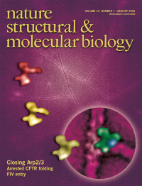
Electron microscopy of the Arp2/3 complex reveals that it exists in multiple conformations, and the closed conformation represents the nucleating form. The EM envelopes of the different conformations (colored shapes) are shown over the raw EM image. pp 26-31