Volume 14
-
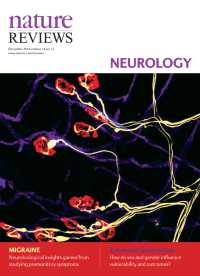
No. 12 December 2018
The image shows lower motor neurons, neuromuscular junctions and vascular plexus of mouse skeletal muscle. Lumbrical muscles of the hindfoot were dissected, and whole-mount immunofluorescent staining was performed before confocal imaging. The neuromuscular and vascular systems can be analysed in mouse models of neurological disorders, such as amyotrophic lateral sclerosis, Charcot–Marie–Tooth disease and spinal muscular atrophy, to enhance our understanding of the underlying neuropathological processes.
-
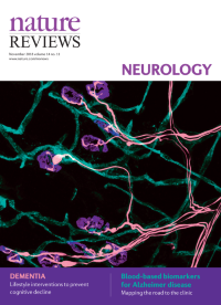
No. 11 November 2018
The image shows lower motor neurons, neuromuscular junctions and vascular plexus of mouse skeletal muscle. Lumbrical muscles of the hindfoot were dissected, and whole-mount immunofluorescent staining was performed before confocal imaging. The neuromuscular and vascular systems can be analysed in mouse models of neurological disorders, such as amyotrophic lateral sclerosis, Charcot–Marie–Tooth disease and spinal muscular atrophy, to enhance our understanding of the underlying neuropathological processes.
-
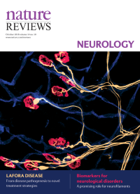
No. 10 October 2018
The image shows lower motor neurons, neuromuscular junctions and vascular plexus of mouse skeletal muscle. Lumbrical muscles of the hindfoot were dissected, and whole-mount immunofluorescent staining was performed before confocal imaging. The neuromuscular and vascular systems can be analysed in mouse models of neurological disorders, such as amyotrophic lateral sclerosis, Charcot–Marie–Tooth disease and spinal muscular atrophy, to enhance our understanding of the underlying neuropathological processes.
-
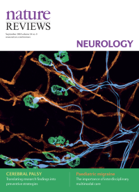
No. 9 September 2018
The image shows lower motor neurons, neuromuscular junctions and vascular plexus of mouse skeletal muscle. Lumbrical muscles of the hindfoot were dissected, and whole-mount immunofluorescent staining was performed before confocal imaging. The neuromuscular and vascular systems can be analysed in mouse models of neurological disorders, such as amyotrophic lateral sclerosis, Charcot–Marie–Tooth disease and spinal muscular atrophy, to enhance our understanding of the underlying neuropathological processes.
-
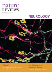
No. 8 August 2018
The image shows lower motor neurons, neuromuscular junctions and vascular plexus of mouse skeletal muscle. Lumbrical muscles of the hindfoot were dissected, and whole-mount immunofluorescent staining was performed before confocal imaging. The neuromuscular and vascular systems can be analysed in mouse models of neurological disorders, such as amyotrophic lateral sclerosis, Charcot–Marie–Tooth disease and spinal muscular atrophy, to enhance our understanding of the underlying neuropathological processes.
-
No. 7 July 2018
The image shows lower motor neurons, neuromuscular junctions and vascular plexus of mouse skeletal muscle. Lumbrical muscles of the hindfoot were dissected, and whole-mount immunofluorescent staining was performed before confocal imaging. The neuromuscular and vascular systems can be analysed in mouse models of neurological disorders, such as amyotrophic lateral sclerosis, Charcot–Marie–Tooth disease and spinal muscular atrophy, to enhance our understanding of the underlying neuropathological processes.
-
No. 6 June 2018
The image shows lower motor neurons, neuromuscular junctions and vascular plexus of mouse skeletal muscle. Lumbrical muscles of the hindfoot were dissected, and whole-mount immunofluorescent staining was performed before confocal imaging. The neuromuscular and vascular systems can be analysed in mouse models of neurological disorders, such as amyotrophic lateral sclerosis, Charcot–Marie–Tooth disease and spinal muscular atrophy, to enhance our understanding of the underlying neuropathological processes.
-
No. 5 May 2018
Cover image supplied by James N. Sleigh, Institute of Neurology, University College London, London, UK. The image shows lower motor neurons, neuromuscular junctions and vascular plexus of mouse skeletal muscle. Lumbrical muscles of the hindfoot were dissected, and whole-mount immunofluorescent staining was performed before confocal imaging. The neuromuscular and vascular systems can be analysed in mouse models of neurological disorders, such as amyotrophic lateral sclerosis, CharcotâMarieâTooth disease and spinal muscular atrophy, to enhance our understanding of the underlying neuropathological processes. Photo copyright James N. Sleigh, supplied by Wellcome Collection (https://wellcomecollection.org/), licensed under CC-BY-NC 4.0 (https://creativecommons.org/licenses/by-nc/4.0/)/colours modified.
-
No. 4 April 2018
Cover image supplied by James N. Sleigh, Institute of Neurology, University College London, London, UK. The image shows lower motor neurons, neuromuscular junctions and vascular plexus of mouse skeletal muscle. Lumbrical muscles of the hindfoot were dissected, and whole-mount immunofluorescent staining was performed before confocal imaging. The neuromuscular and vascular systems can be analysed in mouse models of neurological disorders, such as amyotrophic lateral sclerosis, CharcotâMarieâTooth disease and spinal muscular atrophy, to enhance our understanding of the underlying neuropathological processes. Photo copyright James N. Sleigh, supplied by Wellcome Collection (https://wellcomecollection.org/), licensed under CC-BY-NC 4.0 (https://creativecommons.org/licenses/by-nc/4.0/)/colours modified.
-
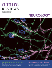
No. 3 March 2018
Cover image supplied by James N. Sleigh, Institute of Neurology, University College London, London, UK. The image shows lower motor neurons, neuromuscular junctions and vascular plexus of mouse skeletal muscle. Lumbrical muscles of the hindfoot were dissected, and whole-mount immunofluorescent staining was performed before confocal imaging. The neuromuscular and vascular systems can be analysed in mouse models of neurological disorders, such as amyotrophic lateral sclerosis, CharcotâMarieâTooth disease and spinal muscular atrophy, to enhance our understanding of the underlying neuropathological processes. Photo copyright James N. Sleigh, supplied by Wellcome Collection (https://wellcomecollection.org/), licensed under CC-BY-NC 4.0 (https://creativecommons.org/licenses/by-nc/4.0/)/colours modified.
-
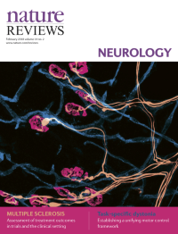
No. 2 February 2018
Cover image supplied by James N. Sleigh, Institute of Neurology, University College London, London, UK. The image shows lower motor neurons, neuromuscular junctions and vascular plexus of mouse skeletal muscle. Lumbrical muscles of the hindfoot were dissected, and whole-mount immunofluorescent staining was performed before confocal imaging. The neuromuscular and vascular systems can be analysed in mouse models of neurological disorders, such as amyotrophic lateral sclerosis, CharcotâMarieâTooth disease and spinal muscular atrophy, to enhance our understanding of the underlying neuropathological processes. Photo copyright James N. Sleigh, supplied by Wellcome Collection (https://wellcomecollection.org/), licensed under CC-BY-NC 4.0 (https://creativecommons.org/licenses/by-nc/4.0/)/colours modified.
-
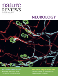
No. 1 January 2018
Cover image supplied by James N. Sleigh, Institute of Neurology, University College London, London, UK. The image shows lower motor neurons, neuromuscular junctions and vascular plexus of mouse skeletal muscle. Lumbrical muscles of the hindfoot were dissected, and whole-mount immunofluorescent staining was performed before confocal imaging. The neuromuscular and vascular systems can be analysed in mouse models of neurological disorders, such as amyotrophic lateral sclerosis, CharcotâMarieâTooth disease and spinal muscular atrophy, to enhance our understanding of the underlying neuropathological processes. Photo copyright James N. Sleigh, supplied by Wellcome Collection (https://wellcomecollection.org/), licensed under CC-BY-NC 4.0 (https://creativecommons.org/licenses/by-nc/4.0/)/colours modified.