Volume 10
-
No. 12 December 2014
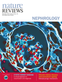
Cover image supplied by Miriam Boersema, University of Groningen, University Medical Center Groningen, Netherlands. Immunofluorescent image of the extracellular matrix in a glomerulus from an allografted rat kidney. The double staining shows the spatial relationship between collagen I and collagen IV. Original lens magnification 40x.
-
No. 11 November 2014
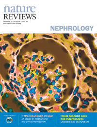
Cover image supplied by Miriam Boersema, University of Groningen, University Medical Center Groningen, Netherlands. Immunofluorescent image of the extracellular matrix in a glomerulus from an allografted rat kidney. The double staining shows the spatial relationship between collagen I and collagen IV. Original lens magnification 40x.
-
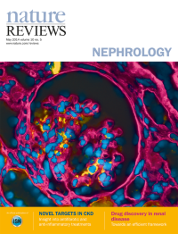
No. 10 October 2014
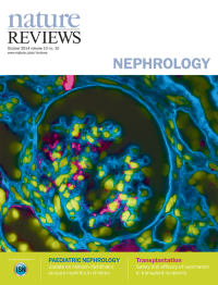
Cover image supplied by Miriam Boersema, University of Groningen, University Medical Center Groningen, Netherlands. Immunofluorescent image of the extracellular matrix in a glomerulus from an allografted rat kidney. The double staining shows the spatial relationship between collagen I and collagen IV. Original lens magnification 40x.
-
No. 9 September 2014
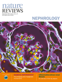
Cover image supplied by Miriam Boersema, University of Groningen, University Medical Center Groningen, Netherlands. Immunofluorescent image of the extracellular matrix in a glomerulus from an allografted rat kidney. The double staining shows the spatial relationship between collagen I and collagen IV. Original lens magnification 40x.
-
No. 8 August 2014
Cover image supplied by Miriam Boersema, University of Groningen, University Medical Center Groningen, Netherlands. Immunofluorescent image of the extracellular matrix in a glomerulus from an allografted rat kidney. The double staining shows the spatial relationship between collagen I and collagen IV. Original lens magnification 40x.
-
No. 7 July 2014
Cover image supplied by Miriam Boersema, University of Groningen, University Medical Center Groningen, Netherlands. Immunofluorescent image of the extracellular matrix in a glomerulus from an allografted rat kidney. The double staining shows the spatial relationship between collagen I and collagen IV. Original lens magnification 40x.
-
No. 6 June 2014
Cover image supplied by Miriam Boersema, University of Groningen, University Medical Center Groningen, Netherlands. Immunofluorescent image of the extracellular matrix in a glomerulus from an allografted rat kidney. The double staining shows the spatial relationship between collagen I and collagen IV. Original lens magnification 40x.
-
No. 5 May 2014
Cover image supplied by Miriam Boersema, University of Groningen, University Medical Center Groningen, Netherlands. Immunofluorescent image of the extracellular matrix in a glomerulus from an allografted rat kidney. The double staining shows the spatial relationship between collagen I and collagen IV. Original lens magnification 40x.
-
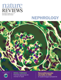
No. 4 April 2014
Cover image supplied by Miriam Boersema, University of Groningen, University Medical Center Groningen, Netherlands. Immunofluorescent image of the extracellular matrix in a glomerulus from an allografted rat kidney. The double staining shows the spatial relationship between collagen I and collagen IV. Original lens magnification 40x.
-
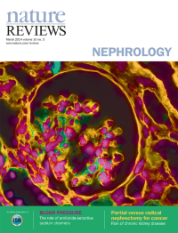
No. 3 March 2014
Cover image supplied by Miriam Boersema, University of Groningen, University Medical Center Groningen, Netherlands. Immunofluorescent image of the extracellular matrix in a glomerulus from an allografted rat kidney. The double staining shows the spatial relationship between collagen I and collagen IV. Original lens magnification 40x.
-
No. 2 February 2014
Cover image supplied by Miriam Boersema, University of Groningen, University Medical Center Groningen, Netherlands. Immunofluorescent image of the extracellular matrix in a glomerulus from an allografted rat kidney. The double staining shows the spatial relationship between collagen I and collagen IV. Original lens magnification 40x.
-
No. 1 January 2014
Cover image supplied by Miriam Boersema, University of Groningen, University Medical Center Groningen, Netherlands. Immunofluorescent image of the extracellular matrix in a glomerulus from an allografted rat kidney. The double staining shows the spatial relationship between collagen I and collagen IV. Original lens magnification 40x.