Volume 14
-
No. 12 December 2017
Cover image supplied by Carolina Tropini, Sonnenburg Group, Stanford University, USA, who is funded by a James S. McDonnell fellowship. Fluorescent in situ hybridization of mouse colon colonized with gnotobiotic microbiota. Tissue was stained by DAPI and the mucus labelled with UEA-1 (Ulex europaeus agglutinin I), bacteria were labelled with fluorescent DNA probes.
-
No. 11 November 2017
Cover image supplied by Carolina Tropini, Sonnenburg Group, Stanford University, USA, who is funded by a James S. McDonnell fellowship. Fluorescent in situ hybridization of mouse colon colonized with gnotobiotic microbiota. Tissue was stained by DAPI and the mucus labelled with UEA-1 (Ulex europaeus agglutinin I), bacteria were labelled with fluorescent DNA probes.
-
No. 10 October 2017
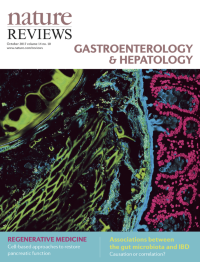
Cover image supplied by Carolina Tropini, Sonnenburg Group, Stanford University, USA, who is funded by a James S. McDonnell fellowship. Fluorescent in situ hybridization of mouse colon colonized with gnotobiotic microbiota. Tissue was stained by DAPI and the mucus labelled with UEA-1 (Ulex europaeus agglutinin I), bacteria were labelled with fluorescent DNA probes.
-
No. 9 September 2017
Cover image supplied by Carolina Tropini, Sonnenburg Group, Stanford University, USA, who is funded by a James S. McDonnell fellowship. Fluorescent in situ hybridization of mouse colon colonized with gnotobiotic microbiota. Tissue was stained by DAPI and the mucus labelled with UEA-1 (Ulex europaeus agglutinin I), bacteria were labelled with fluorescent DNA probes.
-
No. 8 August 2017
Cover image supplied by Carolina Tropini, Sonnenburg Group, Stanford University, USA, who is funded by a James S. McDonnell fellowship. Fluorescent in situ hybridization of mouse colon colonized with gnotobiotic microbiota. Tissue was stained by DAPI and the mucus labelled with UEA-1 (Ulex europaeus agglutinin I), bacteria were labelled with fluorescent DNA probes.
-
No. 7 July 2017
Cover image supplied by Carolina Tropini, Sonnenburg Group, Stanford University, USA, who is funded by a James S. McDonnell fellowship. Fluorescent in situ hybridization of mouse colon colonized with gnotobiotic microbiota. Tissue was stained by DAPI and the mucus labelled with UEA-1 (Ulex europaeus agglutinin I), bacteria were labelled with fluorescent DNA probes.
-
No. 6 June 2017
Cover image supplied by Carolina Tropini, Sonnenburg Group, Stanford University, USA, who is funded by a James S. McDonnell fellowship. Fluorescent in situ hybridization of mouse colon colonized with gnotobiotic microbiota. Tissue was stained by DAPI and the mucus labelled with UEA-1 (Ulex europaeus agglutinin I), bacteria were labelled with fluorescent DNA probes.
-
No. 5 May 2017
Cover image supplied by Carolina Tropini, Sonnenburg Group, Stanford University, USA, who is funded by a James S. McDonnell fellowship. Fluorescent in situ hybridization of mouse colon colonized with gnotobiotic microbiota. Tissue was stained by DAPI and the mucus labelled with UEA-1 (Ulex europaeus agglutinin I), bacteria were labelled with fluorescent DNA probes.
-
No. 4 April 2017
Cover image supplied by Carolina Tropini, Sonnenburg Group, Stanford University, USA, who is funded by a James S. McDonnell fellowship. Fluorescent in situ hybridization of mouse colon colonized with gnotobiotic microbiota. Tissue was stained by DAPI and the mucus labelled with UEA-1 (Ulex europaeus agglutinin I), bacteria were labelled with fluorescent DNA probes.
-
No. 3 March 2017
Cover image supplied by Carolina Tropini, Sonnenburg Group, Stanford University, USA, who is funded by a James S. McDonnell fellowship. Fluorescent in situ hybridization of mouse colon colonized with gnotobiotic microbiota. Tissue was stained by DAPI and the mucus labelled with UEA-1 (Ulex europaeus agglutinin I), bacteria were labelled with fluorescent DNA probes.
-
No. 2 February 2017
Cover image supplied by Carolina Tropini, Sonnenburg Group, Stanford University, USA, who is funded by a James S. McDonnell fellowship. Fluorescent in situ hybridization of mouse colon colonized with gnotobiotic microbiota. Tissue was stained by DAPI and the mucus labelled with UEA-1 (Ulex europaeus agglutinin I), bacteria were labelled with fluorescent DNA probes.
-
No. 1 January 2017
Cover image supplied by Carolina Tropini, Sonnenburg Group, Stanford University, USA, who is funded by a James S. McDonnell fellowship. Fluorescent in situ hybridization of mouse colon colonized with gnotobiotic microbiota. Tissue was stained by DAPI and the mucus labelled with UEA-1 (Ulex europaeus agglutinin I), bacteria were labelled with fluorescent DNA probes.


