Volume 13
-
No. 12 December 2017
This image illustrates the human pancreatic microenvironment in type 2 diabetes mellitus. The pancreatic lobule, which was labelled for CD31 (vasculature) and glucagon (islet), was imaged by deep-tissue confocal microscopy with optical clearing (refractive index: 1.52) to illustrate the fatty infiltration (transmitted light signals) around the endocrine and exocrine pancreas. Image supplied by Shiue-Cheng Tang and Shih-Jung Peng at Department of Medical Science, National Tsing Hua University, Taiwan, and Luc Baeyens and Michael German at Diabetes Center, UCSF, USA.
-
No. 11 November 2017
This image illustrates the human pancreatic microenvironment in type 2 diabetes mellitus. The pancreatic lobule, which was labelled for CD31 (vasculature) and glucagon (islet), was imaged by deep-tissue confocal microscopy with optical clearing (refractive index: 1.52) to illustrate the fatty infiltration (transmitted light signals) around the endocrine and exocrine pancreas. Image supplied by Shiue-Cheng Tang and Shih-Jung Peng at Department of Medical Science, National Tsing Hua University, Taiwan, and Luc Baeyens and Michael German at Diabetes Center, UCSF, USA.
-
No. 10 October 2017
This image illustrates the human pancreatic microenvironment in type 2 diabetes mellitus. The pancreatic lobule, which was labelled for CD31 (vasculature) and glucagon (islet), was imaged by deep-tissue confocal microscopy with optical clearing (refractive index: 1.52) to illustrate the fatty infiltration (transmitted light signals) around the endocrine and exocrine pancreas. Image supplied by Shiue-Cheng Tang and Shih-Jung Peng at Department of Medical Science, National Tsing Hua University, Taiwan, and Luc Baeyens and Michael German at Diabetes Center, UCSF, USA.
-
No. 9 September 2017
This image illustrates the human pancreatic microenvironment in type 2 diabetes mellitus. The pancreatic lobule, which was labelled for CD31 (vasculature) and glucagon (islet), was imaged by deep-tissue confocal microscopy with optical clearing (refractive index: 1.52) to illustrate the fatty infiltration (transmitted light signals) around the endocrine and exocrine pancreas. Image supplied by Shiue-Cheng Tang and Shih-Jung Peng at Department of Medical Science, National Tsing Hua University, Taiwan, and Luc Baeyens and Michael German at Diabetes Center, UCSF, USA.
-
No. 8 August 2017
This image illustrates the human pancreatic microenvironment in type 2 diabetes mellitus. The pancreatic lobule, which was labelled for CD31 (vasculature) and glucagon (islet), was imaged by deep-tissue confocal microscopy with optical clearing (refractive index: 1.52) to illustrate the fatty infiltration (transmitted light signals) around the endocrine and exocrine pancreas. Image supplied by Shiue-Cheng Tang and Shih-Jung Peng at Department of Medical Science, National Tsing Hua University, Taiwan, and Luc Baeyens and Michael German at Diabetes Center, UCSF, USA.
-
No. 7 July 2017
This image illustrates the human pancreatic microenvironment in type 2 diabetes mellitus. The pancreatic lobule, which was labelled for CD31 (vasculature) and glucagon (islet), was imaged by deep-tissue confocal microscopy with optical clearing (refractive index: 1.52) to illustrate the fatty infiltration (transmitted light signals) around the endocrine and exocrine pancreas. Image supplied by Shiue-Cheng Tang and Shih-Jung Peng at Department of Medical Science, National Tsing Hua University, Taiwan, and Luc Baeyens and Michael German at Diabetes Center, UCSF, USA.
-
No. 6 June 2017
This image illustrates the human pancreatic microenvironment in type 2 diabetes mellitus. The pancreatic lobule, which was labelled for CD31 (vasculature) and glucagon (islet), was imaged by deep-tissue confocal microscopy with optical clearing (refractive index: 1.52) to illustrate the fatty infiltration (transmitted light signals) around the endocrine and exocrine pancreas. Image supplied by Shiue-Cheng Tang and Shih-Jung Peng at Department of Medical Science, National Tsing Hua University, Taiwan, and Luc Baeyens and Michael German at Diabetes Center, UCSF, USA.
-
No. 5 May 2017
This image illustrates the human pancreatic microenvironment in type 2 diabetes mellitus. The pancreatic lobule, which was labelled for CD31 (vasculature) and glucagon (islet), was imaged by deep-tissue confocal microscopy with optical clearing (refractive index: 1.52) to illustrate the fatty infiltration (transmitted light signals) around the endocrine and exocrine pancreas. Image supplied by Shiue-Cheng Tang and Shih-Jung Peng at Department of Medical Science, National Tsing Hua University, Taiwan, and Luc Baeyens and Michael German at Diabetes Center, UCSF, USA.
-
No. 4 April 2017
This image illustrates the human pancreatic microenvironment in type 2 diabetes mellitus. The pancreatic lobule, which was labelled for CD31 (vasculature) and glucagon (islet), was imaged by deep-tissue confocal microscopy with optical clearing (refractive index: 1.52) to illustrate the fatty infiltration (transmitted light signals) around the endocrine and exocrine pancreas. Image supplied by Shiue-Cheng Tang and Shih-Jung Peng at Department of Medical Science, National Tsing Hua University, Taiwan, and Luc Baeyens and Michael German at Diabetes Center, UCSF, USA.
-
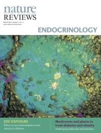
No. 3 March 2017
This image illustrates the human pancreatic microenvironment in type 2 diabetes mellitus. The pancreatic lobule, which was labelled for CD31 (vasculature) and glucagon (islet), was imaged by deep-tissue confocal microscopy with optical clearing (refractive index: 1.52) to illustrate the fatty infiltration (transmitted light signals) around the endocrine and exocrine pancreas. Image supplied by Shiue-Cheng Tang and Shih-Jung Peng at Department of Medical Science, National Tsing Hua University, Taiwan, and Luc Baeyens and Michael German at Diabetes Center, UCSF, USA.
-
No. 2 February 2017
This image illustrates the human pancreatic microenvironment in type 2 diabetes mellitus. The pancreatic lobule, which was labelled for CD31 (vasculature) and glucagon (islet), was imaged by deep-tissue confocal microscopy with optical clearing (refractive index: 1.52) to illustrate the fatty infiltration (transmitted light signals) around the endocrine and exocrine pancreas. Image supplied by Shiue-Cheng Tang and Shih-Jung Peng at Department of Medical Science, National Tsing Hua University, Taiwan, and Luc Baeyens and Michael German at Diabetes Center, UCSF, USA.
-
No. 1 January 2017
This image illustrates the human pancreatic microenvironment in type 2 diabetes mellitus. The pancreatic lobule, which was labelled for CD31 (vasculature) and glucagon (islet), was imaged by deep-tissue confocal microscopy with optical clearing (refractive index: 1.52) to illustrate the fatty infiltration (transmitted light signals) around the endocrine and exocrine pancreas. Image supplied by Shiue-Cheng Tang and Shih-Jung Peng at Department of Medical Science, National Tsing Hua University, Taiwan, and Luc Baeyens and Michael German at Diabetes Center, UCSF, USA.