Volume 15
-
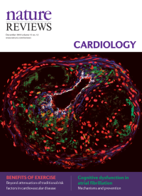
No. 12 December 2018
The picture shows an advanced atherosclerotic lesion in a hypercholesterolaemic mouse, with staining for macrophages, smooth muscle cells, nuclei, and endothelium.
-
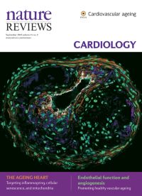
No. 11 November 2018
The picture shows an advanced atherosclerotic lesion in a hypercholesterolaemic mouse, with staining for macrophages, smooth muscle cells, nuclei, and endothelium.
-
No. 10 October 2018
The picture shows an advanced atherosclerotic lesion in a hypercholesterolaemic mouse, with staining for macrophages, smooth muscle cells, nuclei, and endothelium.
-
No. 9 September 2018
The picture shows an advanced atherosclerotic lesion in a hypercholesterolaemic mouse, with staining for macrophages, smooth muscle cells, nuclei, and endothelium.
-
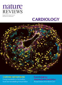
No. 8 August 2018
The picture shows an advanced atherosclerotic lesion in a hypercholesterolaemic mouse, with staining for macrophages, smooth muscle cells, nuclei, and endothelium.
-
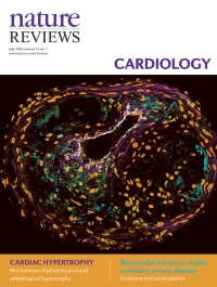
No. 7 July 2018
The picture shows an advanced atherosclerotic lesion in a hypercholesterolaemic mouse, with staining for macrophages, smooth muscle cells, nuclei, and endothelium.
-
No. 6 June 2018
The picture shows an advanced atherosclerotic lesion in a hypercholesterolaemic mouse, with staining for macrophages, smooth muscle cells, nuclei, and endothelium.
-
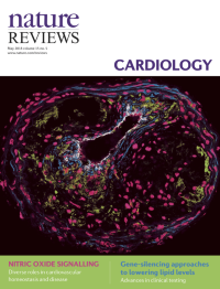
No. 5 May 2018
Cover image supplied by Oliver Soehnlein and Carlos Silvestre-Roig from the Institute for Cardiovascular Prevention (IPEK), Ludwig Maximilian University, Munich, Germany. The picture shows an advanced atherosclerotic lesion in a hypercholesterolaemic mouse, with staining for macrophages, smooth muscle cells, nuclei, and endothelium.
-
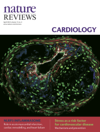
No. 4 April 2018
Cover image supplied by Oliver Soehnlein and Carlos Silvestre-Roig from the Institute for Cardiovascular Prevention (IPEK), Ludwig Maximilian University, Munich, Germany. The picture shows an advanced atherosclerotic lesion in a hypercholesterolaemic mouse, with staining for macrophages, smooth muscle cells, nuclei, and endothelium.
-
No. 3 March 2018
Cover image supplied by Oliver Soehnlein and Carlos Silvestre-Roig from the Institute for Cardiovascular Prevention (IPEK), Ludwig Maximilian University, Munich, Germany. The picture shows an advanced atherosclerotic lesion in a hypercholesterolaemic mouse, with staining for macrophages, smooth muscle cells, nuclei, and endothelium.
-
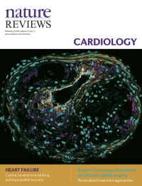
No. 2 February 2018
Cover image supplied by Oliver Soehnlein and Carlos Silvestre-Roig from the Institute for Cardiovascular Prevention (IPEK), Ludwig Maximilian University, Munich, Germany. The picture shows an advanced atherosclerotic lesion in a hypercholesterolaemic mouse, with staining for macrophages, smooth muscle cells, nuclei, and endothelium.
-
No. 1 January 2018
Cover image supplied by Oliver Soehnlein and Carlos Silvestre-Roig from the Institute for Cardiovascular Prevention (IPEK), Ludwig Maximilian University, Munich, Germany. The picture shows an advanced atherosclerotic lesion in a hypercholesterolaemic mouse, with staining for macrophages, smooth muscle cells, nuclei, and endothelium.