Volume 5
-
No. 12 December 2010
Images of slides containing four identical arrays processed with the same hybridoma supernatant. The arrays are composed of whole inactivated influenza viruses and recombinant nucleoproteins. The sameness of the reactivity pattern observed in all array replicas on one slide demonstrates the very low variability among intraslide spots.
-
No. 11 November 2010
Scanning electron micrograph of poly(acrylamide) cryogel showing uniform pore distribution. Original magnification ×40. Image is from the protocol by Kumar and Srivastava on p. 1737.
-
No. 10 October 2010
A confocal micrograph showing direct EGFP epifluorescence in pyramidal neurons of the cerebral cortex taken from a BAC Crym-EGFP P7 transgenic mouse. (from the protocol by Gong, Kus & Heintz)
-
No. 9 September 2010
A Southern blot analyzing terminal restriction fragments in leukocytes. The lanes contain DNA from 32 leukocyte samples along with molecular-weight ladders and, on the far left, DNA from an internal reference sample. Image is from the protocol by Kimura et al.
-
No. 8 August 2010
Chromosome transfer between mature primate oocytes. Chromosomes from metaphase 2arrested oocytes are isolated into membrane- surrounded karyoplasts and placed into the perivetelline space of recipient-enucleated oocytes. Chromosomes are then introduced into these enucleated oocytes via membrane fusion. Image is from the protocol by Tachibana et al. (doi:10.1038/nprot.2010.75).
-
No. 7 July 2010
A monolayer of cultured cells derived from the microvascular endothelium of a human brain. The cells are immunofluorescently stained to show the presence of proteins associated with tight junctions. Image is from the protocol by Bernas et al. on p. 1247. Cover design by Jamel Wooten.
-
No. 6 June 2010
Snapshots from a bioinformatics approach to gene annotation in plants. Individual screens show a typical gene co-expression graph, expression database analysis and microarray analysis used for metabolic pathway prediction. Screen images from the protocol by Tohge and Fernie. Image of monitors from iStockphoto, [|copy|] Milan Zeremski. Cover design by Jamel Wooten.
-
No. 5 May 2010
Purification of anthraquinone derivatives by reverse phase flash column chromatography, from the protocol by Younis Baqi & Christa E Müller.
-
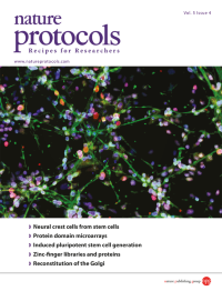
No. 4 April 2010
Specification of human embryonic stem cellneural crest (NC) cells toward peripheral neurons, specifically human pluripotent stem cellderived NC cells differentiated toward sensory neurons (Brn3a+/peripherin+). Image is from the protocol by Lee et al. on p. 688. Cover design by Jamel Wooten.
-
No. 3 March 2010
Mutant (albino) and wild-type zebrafish embryos 48 hours post-fertilization. Image is from the protocol by Mizgirev and Revskoy on p. 383. Cover design by Jamel Wooten.
-
No. 2 February 2010
A composite confocal image of a plucked human hair follicle (lower part; nucleus stained with DAPI and keratinocytes covering the hair with anti-cytokeratin antibody) and a cluster of dopaminergic neurons derived from a plucked hair reprogrammed with Yamanaka factors (upper part stained for Tuj1 and tyrosine hydroxylase). Image is related to the protocol by Aasen and Izpisúa Belmonte.
-
No. 1 January 2010
Growth cone of a primary embryonic mouse motoneuron (E13) cultured for 7 days on laminin 211/221. The neurons were fixed and stained with antibodies against Tau. The picture was taken with a Leica SP2 confocal microscope. Image is from the protocol by Wiese et al, pp 31-38. Cover design by Jamel Wooten.