Thank you for visiting nature.com. You are using a browser version with limited support for CSS. To obtain
the best experience, we recommend you use a more up to date browser (or turn off compatibility mode in
Internet Explorer). In the meantime, to ensure continued support, we are displaying the site without styles
and JavaScript.
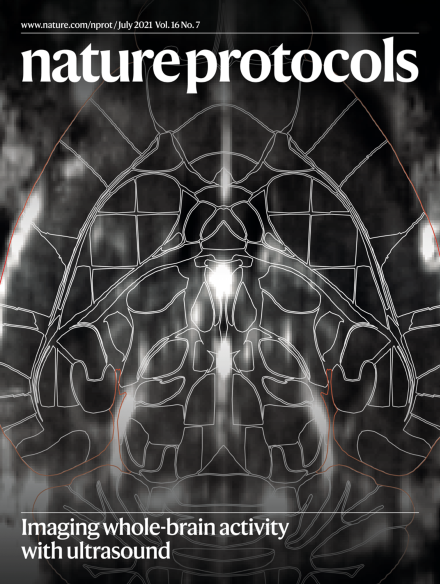
Functional ultrasound imaging (fUS), a hemodynamic-based method, provides a readout of whole-brain activity in awake mice at high spatiotemporal resolution. The image displays a transverse view of the mouse brain microvasculature, captured with fUS. The fUS volume was registered in the Allen Common Coordinate Framework (outlines) using open-source software facilitating the analysis of ~250 brain regions across subjects.
Understanding the heterogeneity of extracellular vesicles is crucial for unraveling their functions. This review describes the benefits, challenges and contributions of the state-of-the art methods used in single-vesicle analysis.
This extension of a previous in vitro digestion protocol provides a subsequent in vitro batch fermentation stage that is carried out afterward to enable investigation of the effect of food on the gut microbiome.
This protocol describes production of lentiviral vectors targeted to receptors present on specific cell types, humanization of mice, administration of the lentiviral vectors and detection of the presence of transduced cell types.
This protocol describes how to perform long-term wide-field imaging of neuronal activity in behaving mice. The procedure discusses how to assemble and calibrate the macroscope, surgical preparation, imaging and data analysis.
To design new multiprotein systems, the authors describe how to combine natural metal-coordinating motifs and hydroxamic acid groups to direct metal-mediated assembly of polyhedral protein architectures and 3D crystalline protein frameworks.
This protocol describes strategies for in situ chemical derivatization and simultaneous quantitative imaging of multiple neurotransmitters and their precursors and metabolites in brain tissue sections using MALDI and desorption electrospray ionization mass spectrometry imaging.
This protocol describes the implantation of Neuropixels probes for chronic recording of neural activity in rats and mice using 3D-printed fixtures. The fixtures enable routine probe reuse, and single-, dual- and movable-probe variants are described.
This approach leverages the rapid, bio-orthogonal inverse electron demand Diels–Alder reaction between a radiolabeled tetrazine and a trans-cyclooctene–bearing antibody to enable pretargeted positron emission tomography imaging and endoradiotherapy in a murine model of cancer.
The hydroperoxide l-tryptophan derivative cis-WOOH regulates vascular tone under inflammatory conditions via redox signaling. This protocol describes the preparation of cis-WOOH and related compounds for use as analytical standards and biochemical tools.
The use of selected ion flow tube mass spectrometry for noninvasive on- or offline analysis of volatile organic compounds within breath is described. This standardized protocol should facilitate noninvasive disease detection and monitoring.
The authors describe a step-by-step workflow for generating genome-wide chromatin interaction maps with nucleosome resolution using proximity ligation and deep sequencing followed by modeling optimal nucleosome position and orientation.
Glycosphingolipids found on the cell membrane play significant roles in many biological processes. This protocol describes how to analyze GSL glycans from cultured cells using a lectin microarray with confirmation by mass spectrometry.
This protocol describes how to set up and use Raman spectroscopy for monitoring the course of solid-state reactions in vibratory ball mills, which will help increase our understanding of the mechanisms and kinetics of mechanochemical reactions.
This protocol for detecting low-copy-number proteins from single cells using plasmonic immunosandwich assays covers fabrication of the plasmonic materials, data collection by Raman scattering and instructions for determining protein concentrations.
Cerebral blood volume dynamics in awake head-fixed mice are tracked via functional ultrasound imaging through a chronic cranial window, giving a high-resolution measure of brain-wide activity.
This protocol describes Deepometry, an open-source application for supervised and weakly supervised deep learning analysis of imaging flow cytometry datasets. The protocol provides runtime scripts for Python, MATLAB and a standalone application.
This protocol uses transient reporters for editing enrichment to facilitate highly efficient base editing of human pluripotent stem cells, enriching for cells edited by adenosine and cytosine base editors and enabling the generation of clonal isogenic human pluripotent stem cell lines.
This protocol describes a computational pipeline for analyzing the single-nucleotide positions and genomic context of DNA-embedded ribonucleotides that have been mapped with any of the currently available ribonucleotide sequencing techniques.
This protocol describes how to identify direct protein targets of noncoding RNAs—Xist, TERRA and U1—and outlines modifications specific to each noncoding RNA and its partners.
This protocol describes how to perform 3D super-resolution imaging on a conventional confocal microscope using metal/graphene-induced energy transfer (MIET/GIET). The protocol covers substrate and sample preparation, imaging and data analysis.
This protocol details how to use Raman spectroscopy for cancer cytopathology diagnosis. Detailed instructions are provided for sample preparation (cervical, oral and lung exfoliated cells), data acquisition, preprocessing and analysis.