Thank you for visiting nature.com. You are using a browser version with limited support for CSS. To obtain
the best experience, we recommend you use a more up to date browser (or turn off compatibility mode in
Internet Explorer). In the meantime, to ensure continued support, we are displaying the site without styles
and JavaScript.
In this protocol extension, the authors detail an in-cell version of their previous in vitro SHAPE-MaP protocol, enabling RNA structure to be probed in living cells.
This protocol describes a semiquantitative glycoproteomics method using sequential treatment with endoglycosidases to create unique mass signatures to determine glycan occupancy and proportion of high-mannose and complex glycans at each glycosite.
VDJ-seq is a DNA-based, high-throughput sequencing method that uses biotinylated J gene primers and UMIs to quantify immunoglobulin diversity in an unbiased manner on a per-cell basis.
CRISPR-EZ achieves 100% delivery of Cas9/sgRNA RNPs by zygote electroporation, enabling efficient incorporation of indels, exon deletions, point mutations, and small insertions into the mouse genome, and outperforming microinjection-based methods.
This protocol describes how to characterize the response properties of individual neurons in mice in vivo using genetically encoded calcium indicators (GECIs), followed by retrieval of the same neurons in brain slices for further analysis in vitro.
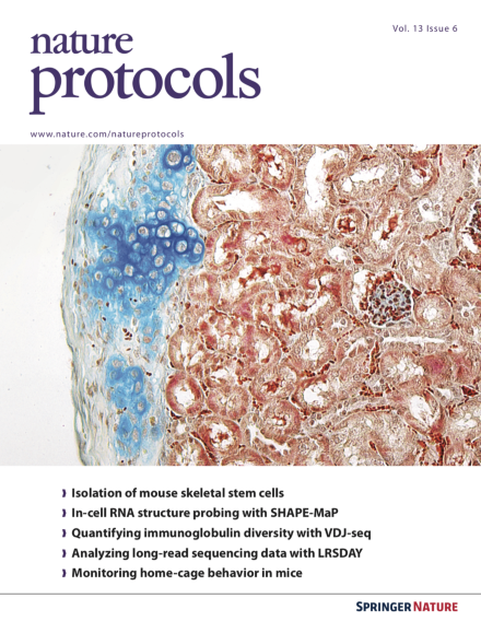
A flow cytometry–based approach using eight surface markers is used to distinguish cells of the skeletal stem cell lineage. Renal subcapsular transplantation and in vitro colony-formation assays are also described for cell characterization.
Many anaerobic microorganisms are difficult to isolate because they grow slowly and rely on interactions with other microorganisms. This protocol describes an approach for the successive enrichment of syntrophic hydrocarbon-degrading microorganisms.
Behavioral phenotyping is often a noisy process in mouse genetics. We developed a home-cage approach controlled by the Phenopy platform, which increases the throughput of data from individual animals and facilitates data sharing across laboratories.
This protocol provides three options for quantifying antibiotic drug concentration in bacterial cells: spectrofluorimetry, microspectrofluorimetry, and kinetics microspectrofluorimetry methods. These approaches are used, respectively, to determine the antibiotic concentration in bacterial populations, in individual cells, and in individual cells across a time course of measurements.
Here, the authors use microsphiltration for the discovery of compounds that stiffen Plasmodium falciparum gametocytes, by filtering compound-exposed gametocytes through microsphere layers in 384-well plates.
A series of diverse in vivo imaging protocols for evaluating hemodynamic response and tissue oxygenation in mouse models of neurological and vascular disorders are described.
Xiao et al. present an approach combining hydrogen–deuterium-exchange mass spectrometry (HDXMS), cross-linking MS (CXMS), and disulfide trapping to elucidate the architecture of protein complexes, using the β2AR-β-arrestin1 complex as an example.
Clustering is an important first step in discovering groups of similar objects in large datasets. ClustEval can be used to find data clusters using multiple algorithms and to evaluate new clustering methods.
This protocol describes how to estimate and spatially resolve the concentration and copy number of fluorescently tagged proteins in live cells using fluorescence imaging and fluorescence correlation spectroscopy (FCS).
This protocol provides a detailed workflow describing how to insert fluorescent markers into all alleles of a gene of interest using CRISPR–Cas9 technology, as well as how to generate and validate homozygous fluorescent knock-in cell lines.
This protocol describes how to use Sclust, a method for copy-number analysis and mutational clustering, to identify subclonal populations in tumor samples.