Thank you for visiting nature.com. You are using a browser version with limited support for CSS. To obtain
the best experience, we recommend you use a more up to date browser (or turn off compatibility mode in
Internet Explorer). In the meantime, to ensure continued support, we are displaying the site without styles
and JavaScript.
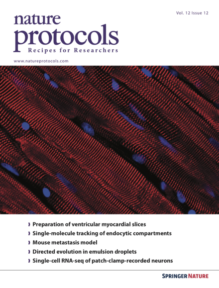
Watson et al. provide a protocol to study cardiac structure using slices of living myocardium from large and small mammals. Shown here is an example of immunohistochemical staining by confocal microscopy of a slice prepared from a rat left ventricle. The sarcomeric apparatus of the cardiac myocytes is labeled with α-actinin (red) and the nuclei of myocytes and non-myocytes with Hoechst 33342 (blue). Image taken from the protocol by Watson et al. doi:10.1038/nprot.2017.139. Cover design by Jamel Wooten.
Here the authors provide an extension to their earlier RNA interactome capture protocol. This Protocol Extension describes RBDmap—a method to identify the regions of RNA-binding proteins engaged in native interactions with RNA, in a proteome-wide manner.
In this protocol, the authors present an experimental metastasis assay in which cancer cells are injected into the tail vein of a mouse, and the resulting secondary organ colonization is assessed, primarily in the lung, 10 d later.
This protocol describes how to use ChromHMM, a robust open-source software package that enables the learning of chromatin states, annotates their occurrences across the genome, and facilitates their biological interpretation.
This protocol describes the procedures for compartmentalized partnered replication (CPR), an emulsion-based directed evolution method for the generation of proteins, genetic elements, and genetic circuits with improved or altered function.
This protocol describes the generation of mice entirely derived from genome-edited embryonic stem cells, enabling the production of transgenic mice in a single generation.
This protocol describes how to integrate whole-cell patch-clamp in single neurons from mouse brain tissue slices with single-cell RNA sequencing and morphological recovery.
This protocol describes how to chemically synthesize membrane proteins through the installation of solubilizing removable backbone modification tags into hydrophobic transmembrane peptides. The implementation of the protocol is demonstrated by the chemical synthesis of phosphorylated M2 (M2-pSer64), a 97-aa proton channel protein from the influenza A virus. The synthesis of M2-pSer64 at milligram scale takes ∼200 working hours (excluding the time for lyophilizations).
Protein crystallization still presents a challenge for X-ray crystallography. This protocol describes the Langmuir–Blodgett nanotemplate method, in which 2D protein LB nanotemplates trigger formation of 3D protein crystals by hanging-drop vapor diffusion.
This protocol describes a pulse–chase approach to studying activity-dependent internalization of fluorescent ligands into endocytic compartments using subdiffractional single-particle tracking in live hippocampal neurons.
This protocol describes how to obtain 100- to 400-μm-thick slices of a living myocardium from rodents, pigs, humans and dogs that retain the native multicellularity, architecture and physiology of the heart.
In this protocol, the authors provide a strategy and set of methods to analyze restriction-site-associated DNA-sequencing (RAD-seq) data using Stacks, enabling the genome-wide discovery and genotyping of SNPs across a range of systems.