Thank you for visiting nature.com. You are using a browser version with limited support for CSS. To obtain
the best experience, we recommend you use a more up to date browser (or turn off compatibility mode in
Internet Explorer). In the meantime, to ensure continued support, we are displaying the site without styles
and JavaScript.
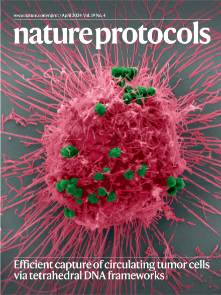
Tumor cell captured via magnetic beads functionalized with tetrahedral DNA frameworks
Scanning electron micrograph of streptavidin-labeled magnetic beads (green) bound to biotin-labeled tetrahedral DNA frameworks anchored with aptamers to a HepG2 cell (pink). See Chen, Y. et al.
Image: [ Yirong Chen, Dekai Ye and Min Li, Shanghai Jiao Tong University] Cover design: S. Whitham
The controlled positioning and stoichiometry of aptamers on tetrahedral DNA frameworks enables the synthesis of targeted probes for the capture of circulating tumor cells from blood samples.
Light-activated assembly of split-protein fragment pairs using the covalent SpyTag/SpyCatcher peptide–protein reaction can be used to modulate biological protein activity in solution, biomaterials and cells.
A light sheet fluorescence microscopy approach for the analysis of immune cell composition, vascularization, tissue perfusion and hypoxia enables spatial imaging of the tissue microenvironment in cancer and cardiovascular diseases.
This protocol provides detailed guidelines for using rMATS-turbo—the latest implementation of the popular software for the discovery and quantification of alternative splicing events from RNA sequencing data—exemplified in two representative scenarios.
A web-based tool to guide the lead optimization process by improving calculation of substructure modifications of candidate compounds with improved absorption, distribution, metabolism, excretion, and toxicity profiles.
A detailed workflow covering 3D pathology, including tissue preparation, imaging with light-sheet fluorescence microscopy, tools for initial data processing in Python (e.g., stitching, intensity leveling and false coloring) and data quality control.
This protocol describes an efficient method to reconstitute the onset of gametogenesis by co-culturing resetting hPGCLCs with human hindgut organoids, providing a new framework to clarify the physiological and pathological crosstalk between hPGCs and the hindgut.
RNA-binding proteins orchestrate many aspects of plant development and environmental responses. This protocol describes an optimized plant individual-nucleotide-resolution cross-linking and immunoprecipitation method for genome-wide identification of RNA-binding protein binding sites on their cognate RNAs at single-nucleotide resolution.
The identification and quantitative characterization of tau posttranslational modifications in brain tissue using mass spectrometry provide a comprehensive and untargeted approach to profiling pathological tau in neurodegenerative diseases.
The formation and functional relevance of N6-methyladenosine sites are key unanswered questions in the field of RNA biology. The protocol describes an unbiased sequencing-based method for the characterization of the global distribution and stoichiometry of N6-methyladenosine sites.