Volume 6
-
No. 12 December 2009
This photograph of an Arabidopsis thaliana (thale cress) anther took first place in the 2009 Nikon Small World photomicrography competition. The image was taken by Heiti Paves of Tallinn University of Technology in Tallinn, Estonia using a confocal microscope at ×20 magnification. Other images from this year's competition are on display at http://www.nikonsmallworld.com/.
-
No. 11 November 2009
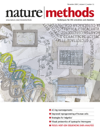
An artistic interpretation of the sequencing process from the DNA molecule to the decoded bases. Cover by Erin Dewalt.
-
No. 10 October 2009
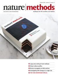
This delicious version of Nature Methods' inaugural issue was baked by How Sweet It Is (http://www.howsweetitispastry.com/), New York. Labeling of gangliosides in cellular membrane and nucleus is in red and blue fondant, respectively. Photography by Christina Holmes. Special 5th anniversary issue feature starts on p701.
-
No. 9 September 2009
A three-dimensional digital atlas of Caenorhabditis elegans with single-cell resolution. Cover art by Hanchuan Peng. Article p667
-
No. 8 August 2009
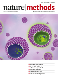
Artistic rendition of human cytomegalovirus particles containing either a fluorescent marker protein stabilized by a ligand or a degraded marker protein in the absence of ligand. The cartoon is based on a method described in the Brief Communication on p577 in which the stabilizing ligand was applied to cytomegalovirus immediate early proteins. Cover design by Erin Dewalt.
-
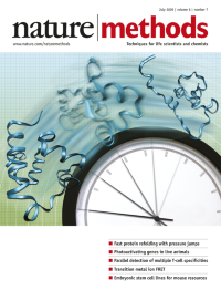
No. 7 July 2009
The 'speed limit' of protein refolding is attained with a method to induce large, sub-microsecond jumps in pressure. Cover by Erin Dewalt, based on images from Martin Gruebele. Article p515, News and Views p490
-
No. 6 June 2009
Fly image from http://www.sxc.hu/, adapted by Erin Boyle. Articles p451, p458, Brief Communications p431, p435, p439, News and Views p413
-
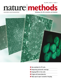
No. 5 May 2009
Artistic rendering by Erin Boyle of brightfield micrographs showing an emerging human induced pluripotent stem (iPS) cell colony (provided by Akitsu Hotta and James Ellis) and a fluorescence micrograph showing chimeric mouse embryos made using mouse iPS cells (provided by Kosuke Yusa and Allan Bradley). Articles, p363, p370
-
No. 4 April 2009
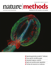
A new method, hopping probe ion conductance microscopy, can be used to discern topological features like stereocilia on live mouse auditory hair cells. Cover by Erin Boyle, based on images provided by Pavel Novak. Brief Communication, p279
-
No. 3 March 2009
Sialic acidcontaining glycoproteins on mammalian cells can be tagged with fluorophores via an efficient chemical labeling approach. Cover design by Erin Boyle. Brief Communication, p207
-
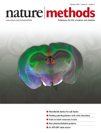
No. 2 February 2009
A section of a mouse brain labeled with multiple transsynaptic pseudorabies viruses expressing different colored proteins. Cover design by Erin Boyle based on an image provided by Botond Roska. Brief Communication, p127
-
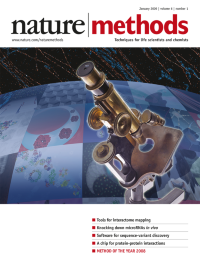
No. 1 January 2009
Super-resolution fluorescence microscopy is likely to revolutionize our understanding of cellular biology and is Nature Methods' pick for Method of the Year 2008. Cover design by Erin Boyle. Special feature starts on p15.